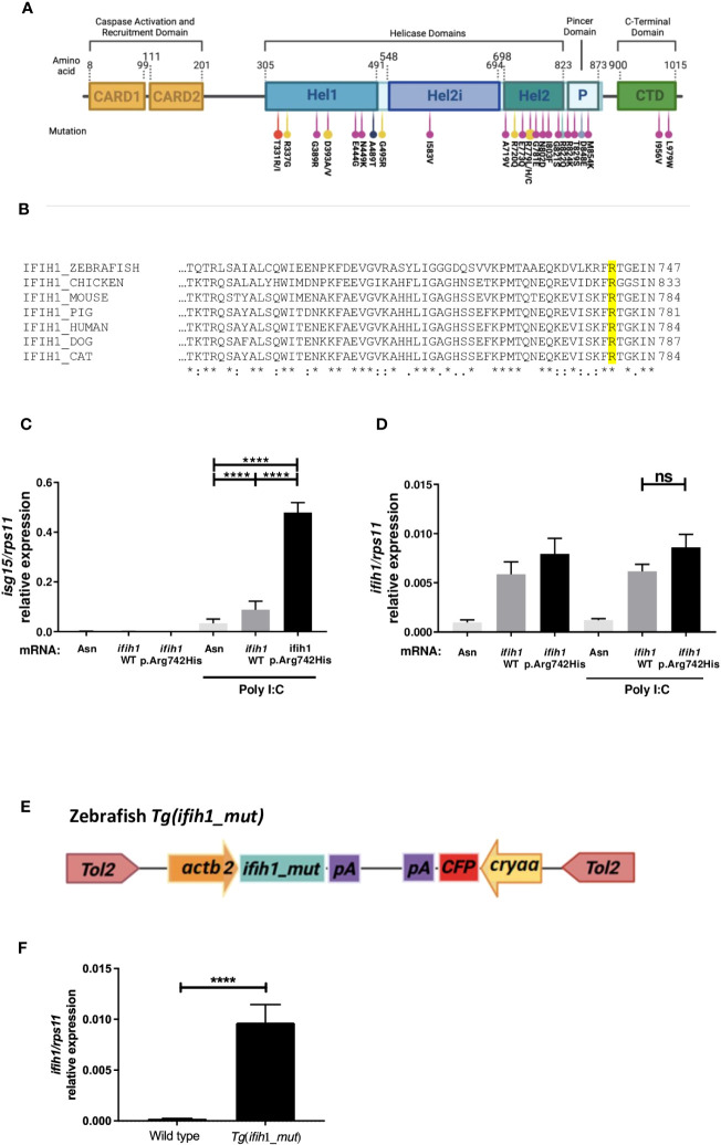
IMAGE

Figure 1

ID
ZDB-IMAGE-231212-29
Source
Figures for Bernal-Bermúdez et al., 2023
Image
Figure Caption

Figure 1

Localization, sequence alignments, ifih1 mRNA overexpression, and Tg(ifih1_mut) line design. (A) Protein diagram showing the positions of protein domains and their amino acid boundaries within the 1,025 residues of IFIH1 (MDA5) protein. The 28 previously published mutations are annotated. CARD, caspase activation recruitment domain; Hel, helicase domain, where Hel1 and Hel2 are the two conserved core helicase domains and Hel2i is an insertion domain that is conserved in the RIG-I–like helicase family; P, pincer or bridge region connecting Hel2 to the C-terminal domain (CTD) involved in binding double-stranded RNA. Red (24), yellow (2), purple (22), black (16), blue (4), and light purple (23). (B) Protein sequence alignment of IFIH1 from different species; IFIH1_ZEBRAFISH (ENSDART00000003913.10), IFIH1_CHICKEN (ENSGALT00010050768.1), IFIH1_MOUSE (ENSMUST00000028259.12), IFH1_PIG (ENSSSCT00000061806.3), IFIH1_HUMAN (ENST00000649979.2), IFH1_DOG (ENSCAFT00845053824.1), IFIH1_CAT (ENSFCAT00000026735.4). (C, D) RT-qPCR of isg15(C) or ifih1(D) from 3-dpf zebrafish larvae injected with wild-type (WT) or mutant (p.Arg742His) ifih1 mRNA with or without 25 pg/egg of poly I:C. (E) Scheme showing zebrafish p.Arg742His transgenic (Tg) design Tg(ifih1_mut) and (F) expression of ifih1 in the transgenic line Tg(ifih1_mut) by RT-PCR. p-values were calculated using one-way ANOVA and Tukey multiple range test. ns, not significant; ****p ≤ 0.0001.

Acknowledgments
This image is the copyrighted work of the attributed author or publisher, and ZFIN has permission only to display this image to its users. Additional permissions should be obtained from the applicable author or publisher of the image. Full text @ Front Immunol