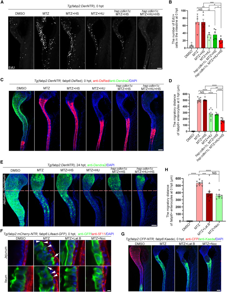
Fig. 3 Cell division and filopodia/lamellipodia are essential for ileal enterocyte migration and jejunal regeneration (A) During the MTZ-induced injury, incubation with hydroxyurea (HU) or overexpression of cdkn1c alone, or HU treatment together with cdkn1c overexpression, significantly reduced the cell proliferation. (B) Statistics showing the number of EdU+ cells in the intestine (n = 6). Data are presented as mean ± SEM. ∗p < 0.05, ∗∗p < 0.01, ∗∗∗p < 0.001, two-tailed unpaired t test; NS, not significant. (C and D) During the MTZ-induced injury, incubation with HU or overexpression of cdkn1c alone, or HU treatment together with cdkn1c overexpression, significantly reduced the migration distance of ileal enterocytes (C), which was validated by the statistical analyses (D, n = 8). Data are presented as mean ± SEM. ∗∗∗p < 0.01, ∗∗∗∗p < 0.0001, two-tailed unpaired t test; NS, not significant. (E) Jejunal regeneration was significantly impaired by incubating with HU or overexpression of cdkn1c alone, or HU treatment together with cdkn1c overexpression from MTZ treatment to 24 hpt. (F–H) Migratory ileal enterocytes formed filopodia (white arrowheads) and lamellipodia (white arrows). The microfilament inhibitor Lat.B and microtube inhibitor Noc blocked the formation of lamellipodia and filopodia (F), then repressed the ileal enterocyte migration (G), which was validated by the statistical analyses (H, n = 8). Data are presented as mean ± SEM. ∗∗∗p < 0.001, ∗∗∗∗p < 0.0001, two-tailed unpaired t test; NS, not significant. Lat.B, latrunculin B; Noc, nocodazole; HS, heat shock. Scale bars, 5 μm (F) and 50 μm (A, C, E, and G). See also Figure S3.
Image
Figure Caption
Acknowledgments
This image is the copyrighted work of the attributed author or publisher, and
ZFIN has permission only to display this image to its users.
Additional permissions should be obtained from the applicable author or publisher of the image.
Full text @ Cell Rep.

