IMAGE

Fig. 1.

ID
ZDB-IMAGE-231204-49
Source
Figures for Mao et al., 2023
Image
Figure Caption

Fig. 1.

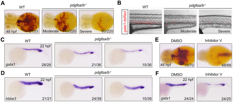
Suppression of PDGF signaling leads to a reduction of erythrocytes. (A) Detection of hemoglobin levels by o-dianisidine staining in wild-type (WT) and pdgfαa−/−;pdgfαb−/− (pdgfαa/b−/−) embryos at 48 hpf. Ventral view. (B) Merged brightfield and confocal images from live imaging of pdgfαa−/−;pdgfαb−/− embryos in the Tg(gata1:dsRed) background at 32 hpf. Lateral view with anterior to the left. Scale bar: 100 μm. (C,D) Expression analysis of (C) gata1 and (D) hbbe3 in WT and pdgfαa−/−;pdgfαb−/− embryos at 22 hpf by in situ hybridization. Lateral view with anterior to the left. (E,F) WT embryos were treated with 0.25 μM inhibitor V from shield stage then harvested at the indicated developmental stages for (E) o-dianisidine staining (ventral view) or (F) gata1 in situ hybridization (lateral view with anterior to the left). Images shown are representative of the observed phenotypes. In A and C-F, the number of embryos displaying each phenotype out of the total number assayed is indicated. Images in B are representative of 35 WT embryos and 40 pdgfαa/b−/− embryos. The number of red blood cells was moderately decreased in about two-thirds of the pdgfαa/b−/− embryos, and severely reduced in the rest of the mutant embryos.

Acknowledgments
This image is the copyrighted work of the attributed author or publisher, and ZFIN has permission only to display this image to its users. Additional permissions should be obtained from the applicable author or publisher of the image. Full text @ Development