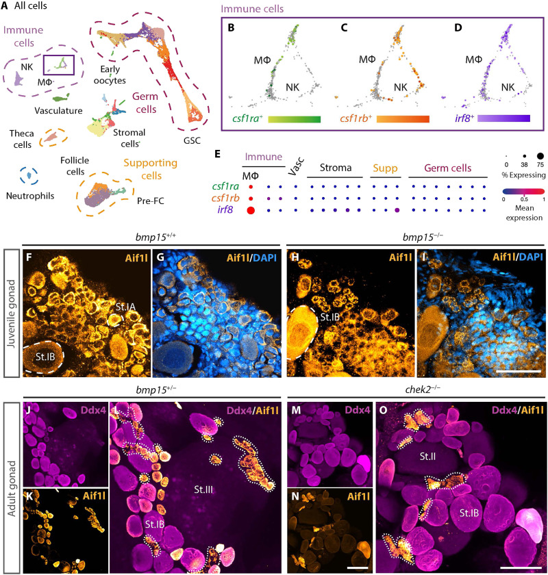
Fig. 2.
Macrophages are resident ovary cells in juvenile and adult zebrafish ovaries.
(A) UMAP plot of cell types present in 40-dpf ovary. (B to D) Magnified view of populations boxed in (A) showing expression of indicated genes. MΦ, macrophages; NK, natural killer–like cells; FC, follicle cells; GSC, germline stem cells; Vasc, vasculature. (E) Analysis of indicated macrophage gene expression profiles in ovarian clusters represented by dot plot graph. (F to O) Immunostained (F) to (I) juvenile and (J) to (O) adult gonads of indicated genotypes. Aif1l (yellow) labels macrophages, Ddx4 (magenta) labels germ cells, and DAPI (blue) labels DNA. Dotted lines mark macrophages. Scale bars, (F) to (I) 50 μm and (J) to (L) 100 μm. St.II, stage II oocyte (apparent cortical alveoli and a vitelline envelope); St.III, stage III oocyte (increased size and apparent yolk).
Acknowledgments
This image is the copyrighted work of the attributed author or publisher, and
ZFIN has permission only to display this image to its users.
Additional permissions should be obtained from the applicable author or publisher of the image.
Full text @ Sci Adv
Your Input Welcome
Thank you for submitting comments. Your input has been emailed to ZFIN curators who may contact you if
additional information is required.
Oops. Something went wrong. Please try again later.