
Fig. 1
- ID
- ZDB-IMAGE-231121-60
- Publication
- Zhang et al., 2023 - Novel biallelic variants in the PLEC gene are associated with severe hearing loss
- All Figures
- Figures for Zhang et al., 2023
|
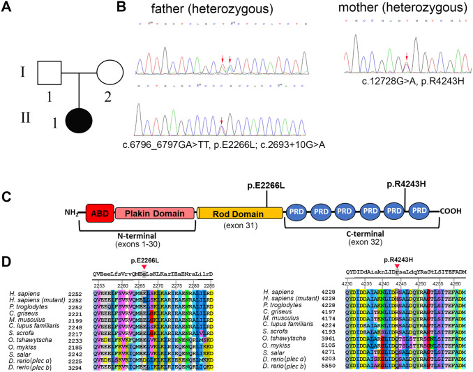
Fig. 1 Pedigree and gene analysis of PLEC in a pediatric patient with ANSD (Family #1). A: Pedigree of the affected family member with ANSD. B: Trio whole exome sequencing, biallelic PLEC variants segregating in family #1 were confirmed by Sanger sequencing. C: Schematic representation of the plectin protein with the newly identified mutations. The central a-helical rod domain, which can form a coiled-coil structure after dimerization, links the N- and C-terminal globular domains. The plakin domain consists of 9 spectrin repeats. ABD: actin binding domain, PRD: plectin repeat domain. D: Amino acid alignment of the regions containing the plectin mutations. Capital letters at the top denote conserved amino acids among all the species.
Reprinted from Hearing Research, 436, Zhang, T., Xu, Z., Zheng, D., Wang, X., He, J., Zhang, L., Zallocchi, M., Novel biallelic variants in the PLEC gene are associated with severe hearing loss, 108831, Copyright (2023) with permission from Elsevier. Full text @ Hear. Res.