IMAGE

Fig. 4

ID
ZDB-IMAGE-231121-44
Source
Figures for Teske et al., 2023
Image
Figure Caption

Fig. 4

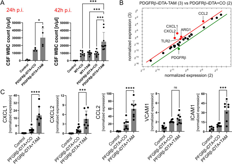
Cerebrospinal (CSF) white blood cell (WBC) counts at 24 and 42 h post-infection in TAM-treated PDGFRB::creER2-iDTA mice (PDGFRβ-iDTA) that are largely missing pericytes compared to control groups (A). Prime™ PCR array analysis of cDNA obtained from brains of infected transgenic mice revealed higher expression levels of CXCL1, CXCL2, and CCL2 (and also of TLR2 and ARG1), but lower PDGFRβ expression following pericyte depletion (B). The array results were confirmed by supplemental RT-PCR analyses (C), which showed increased brain expression of CXCL1, CXCL2, and CCL2 in TAM-treated PDGFRB::creER2-iDTA mice. Data are given as individual values as well as means ± SD. *P < 0.05, **P < 0.01, ***P < 0.001, ****P < 0.0001, using ANOVA with Tukey’s multiple comparisons test

Acknowledgments
This image is the copyrighted work of the attributed author or publisher, and ZFIN has permission only to display this image to its users. Additional permissions should be obtained from the applicable author or publisher of the image. Full text @ J Neuroinflammation