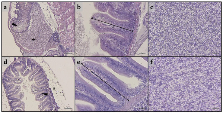
Example of histomorphology of intestine and liver parenchyma of zebrafish (a–c) larvae and (d–f) juveniles from the present study. (a) Representative section of liver (*) and intestine (arrowhead) of a whole-embedded zebrafish larva fed CTRL diet; (b) details of mucosal folds of intestine from a zebrafish larva fed PG diet (double-headed arrow indicate mucosal folds height); (c) liver parenchyma from a zebrafish larva fed A1+ diet; (d) section of intestine (arrowhead) from a zebrafish juvenile fed A2+ diet (# indicate perivisceral adipose tissue); (e) details of mucosal folds of intestine from a zebrafish juvenile fed ROT diet (double-headed arrow indicate mucosal folds height); (f) liver parenchyma from a zebrafish juvenile fed A− diet. Scale bars: a = 200 μm; b,c,e,f = 20 μm; d = 100 μm.
Acknowledgments
This image is the copyrighted work of the attributed author or publisher, and
ZFIN has permission only to display this image to its users.
Additional permissions should be obtained from the applicable author or publisher of the image.
Full text @ Animals (Basel)
Your Input Welcome
Thank you for submitting comments. Your input has been emailed to ZFIN curators who may contact you if
additional information is required.
Oops. Something went wrong. Please try again later.