
Image
|
Figure Caption
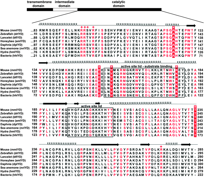
Fig. 3 Multiple sequence alignment of the catalytic domains of IYD homologs. Alignment of homologs chosen for protein expression was generated using MUSCLE.45 Numbering of the amino acids for each protein is indicated on the left and right of the alignment. Residues in red (or white) are fully conserved, and the sequences forming the active site lids are indicated with a box. Key residues coordinating to substrate and FMN are indicated with an (*) and (+), respectively. Secondary structure elements are derived from the crystal structure of mmIYD bound to DIT (PDB ID 3GH8).11
Acknowledgments
This image is the copyrighted work of the attributed author or publisher, and
ZFIN has permission only to display this image to its users.
Additional permissions should be obtained from the applicable author or publisher of the image.
Full text @ Mol. Biosyst.