
Image
|
Figure Caption
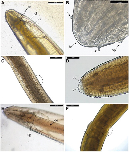
Fig. 2 Photomicrographs of an Eustrongylides spp. larvae (A). Cephalic extremity displaying neural rings (nr), cuticle layer (cl), and part of a vascular system (vs); (B) inner papillae (ip, dotted arrows) and outer papillae (op, arrow); (C) medial part of the larva body and detail of transversal striations; (D) caudal extremity of a female larva presenting the vulva (v) and immature circular reproductive structure (*) and anal canal (ac); (E) anterior extremity presenting cephalic glands (cg) and detail of the esophagus lumen (oe); (F) esophagus–intestinal transition.
Acknowledgments
This image is the copyrighted work of the attributed author or publisher, and
ZFIN has permission only to display this image to its users.
Additional permissions should be obtained from the applicable author or publisher of the image.
Full text @ Zebrafish