
Fig. 1
- ID
- ZDB-IMAGE-231102-51
- Publication
- Sun et al., 2023 - Exposure to PFOA and its novel analogs disrupts lipid metabolism in zebrafish
- All Figures
- Figures for Sun et al., 2023
|
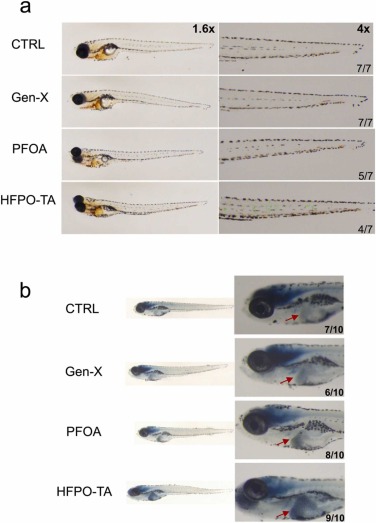
Fig. 1 Representative images of O-Dianisidine staining (a) and Sudan black B staining (b) in zebrafish after 72 hpe exposure to PFOA and alternatives. O-Dianisidine stains mature erythrocyte to reflect abnormalities in the circulatory system, and the green arrows and circles indicates a positive stain, which is dark red. Among them, PFOA, Gen-X was relatively normal, while the HFPO-TA group showed significant erythrocyte retention. n = 7, the lower right corner indicates the number of cases with the same representative image. Sudan black B staining was mainly observed for neutrophil aggregation. The figure shows the aggregation of neutrophils in all three exposure groups, and the red arrows indicate the aggregation in the liver region. n = 10, the lower right corner indicates the number of cases with the same representative image.