IMAGE

Fig 2

ID
ZDB-IMAGE-231101-22
Source
Figures for Chang et al., 2023
Image
Figure Caption

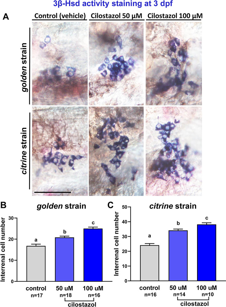
Fig 2 The effect of cilostazol on the morphology and cell number of steroidogenic interrenal tissue in the zebrafish embryo at 3 dpf.

The embryos of golden and citrine strains were treated with cilostazol-supplemented egg water at 50 and 100 μM respectively, or with the vehicle control. The treated embryos were harvested at 3 dpf for whole-mount 3βHsd activity staining. (A) The stained interrenal cells (bluish purple signals at the cytoplasm) were detected on the ventral surface of deyolked embryos with anterior oriented to the left, by using Nomarski microscopy. Scale bar, 50 μm. The number of cells positive for 3βHsd activity staining in the treated golden and citrine embryos is quantified in (B) and (C) respectively. Sample images for the ImageJ quantification of interrenal cells are shown in S1 Fig. Histograms with different letters above them are significantly different (ANOVA and Duncan’s multiple test, P < 0.05).

Acknowledgments
This image is the copyrighted work of the attributed author or publisher, and ZFIN has permission only to display this image to its users. Additional permissions should be obtained from the applicable author or publisher of the image. Full text @ PLoS One