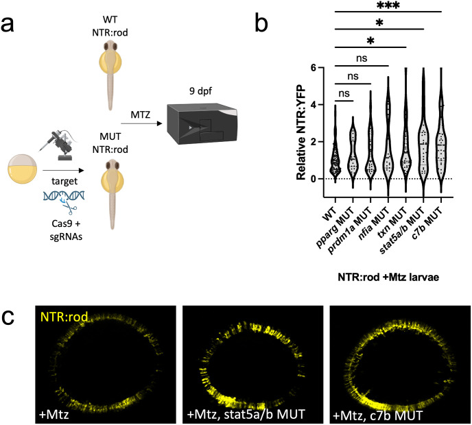
IMAGE

Fig 7

ID
ZDB-IMAGE-231024-28
Source
Figures for Emmerich et al., 2023
Image
Figure Caption

Fig 7 Rod cell regeneration is enhanced in txn, stat5a/b and c7b crispants.

(a) Experimental design depicting CRISPR/Cas9-based targeting of target genes via CRISPR/Cas9 in NTR-rod embryos. Control (+Mtz) and mutated NTR-rod larvae (+Mtz, gene target MUT) were treated with 10mM Mtz from 5–6 dpf and rod fluorescence was assessed via plate reader assay at 9 dpf (to examine the extent of rod cell regeneration). Created with Biorender.com. (b) Quantification of rod cell regeneration by plate reader assay at 9 dpf in Mtz-treated control and MUT larvae for target genes pparg, prdm1a, nfia, txn, stat5a/b and c7b. (c) Representative in vivo confocal images of regenerated rod cells at 9 dpf in control (+Mtz) and stat5a/b or c7b MUT larvae. For statistical comparisons, Welch’s one-way ANOVA was followed by student’s t test with Dunnett’s method for multiple comparisons correction. Asterisks indicate the following p-value ranges: * = p<0.05, ** = p<0.01, *** = p<0.001, and **** = p<0.0001, “ns” indicates p>0.05.

Acknowledgments
This image is the copyrighted work of the attributed author or publisher, and ZFIN has permission only to display this image to its users. Additional permissions should be obtained from the applicable author or publisher of the image. Full text @ PLoS Genet.