IMAGE

Figure 6.

ID
ZDB-IMAGE-231019-11
Source
Figures for Torraca et al., 2023
Image
Figure Caption

Figure 6.

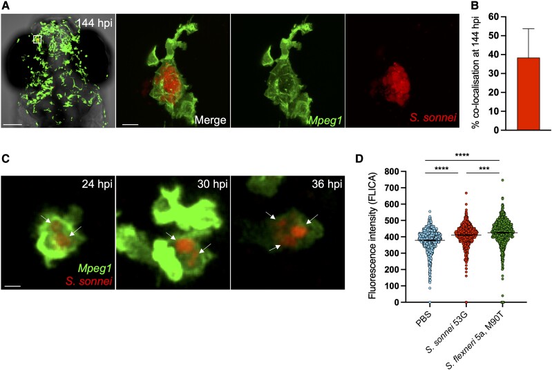
Shigella can establish persistent infection of macrophages in vivo. A, Head region and individual macrophage detail from a representative S. sonnei–infected zebrafish larva at 144 hpi. Tg(mpeg1::Gal4-FF)gl25/Tg(UAS::LIFEACT-GFP)mu271 larvae (Mpeg1, with macrophages in green) were injected at 3 dpf in the hindbrain ventricle with 1000 CFU of mCherry-labeled S. sonnei 53G (red). An infected macrophage harboring persistent bacteria is magnified. Scale bars: 100 μm, left; 10 μm, right and inset. B, At the persistent infection stage, 40% of bacterial fluorescence colocalizes with mpeg1+ macrophages. C, Individual Tg(mpeg1::Gal4-FF)gl25/Tg(UAS::LIFEACT-GFP)mu271 macrophage (Mpeg1, green) harboring mCherry–S. sonnei (red, indicated by arrows) followed for 12 hours (from 24 to 36 hpi). At 2 dpf, 1000 CFU of bacteria were delivered systemically in larvae. Scale bar: 10 μm. D, As compared with S. flexneri M90T, S. sonnei 53G induces a reduced level of caspase 1 activation in vivo in the zebrafish model. At 3 dpf, 10000 CFU of bacteria were delivered systemically in larvae. Data: mean ± SEM. Statistics: 1-way analysis of variance with Sidak correction. ***P < .001. ****P < 0.0001. CFU, colony-forming unit; dpf, days postfertilization; hpi, hours postinfection; PBS, phosphate-buffered saline.

Acknowledgments
This image is the copyrighted work of the attributed author or publisher, and ZFIN has permission only to display this image to its users. Additional permissions should be obtained from the applicable author or publisher of the image. Full text @ J. Infect. Dis.