IMAGE

Fig. 1

ID
ZDB-IMAGE-231002-39
Source
Figures for Yao et al., 2023
Image
Figure Caption

Fig. 1

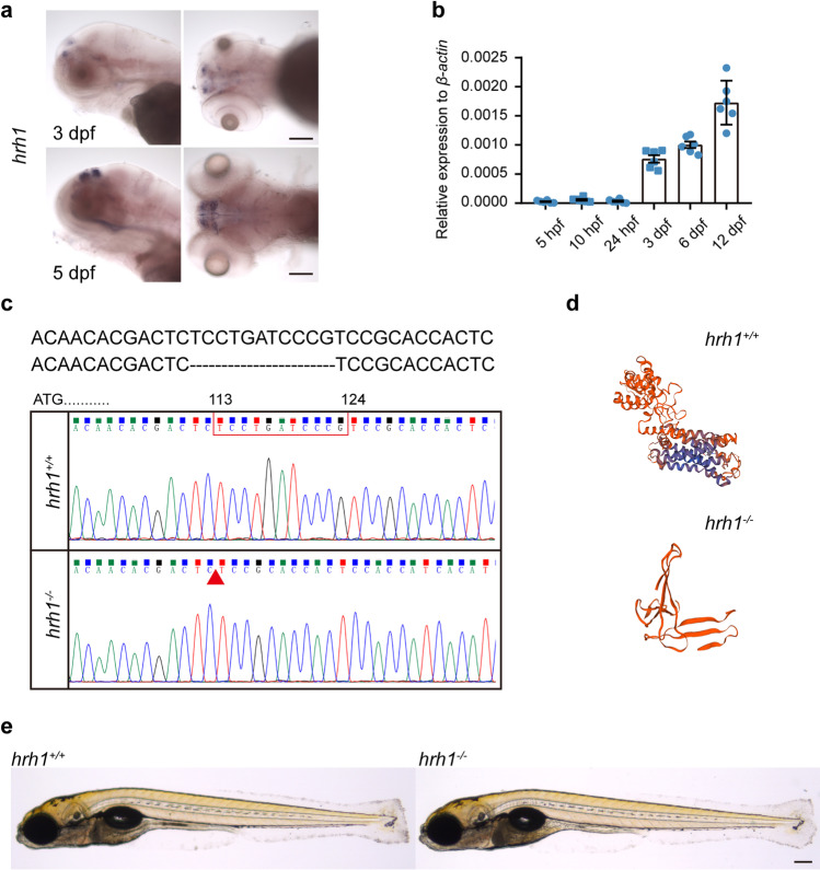
hrh1 expression in Turku wt fishes and the mutation site of hrh1−/− fish. a In situ hybridization of hrh1 in Turku wt zebrafish at 3 dpf (lateral and dorsal view), 5 dpf (lateral and dorsal view). Scale bars: 50 µm. b qRT-PCR results of hrh1 mRNA expression at 5 hpf, 10 hpf, 24 hpf, 3 dpf, 6 dpf, and 12 dpf. N = 6. Mean ± SEM. c The mutation site of hrh1/ zebrafish and genomic DNA sequencing results. d Predicated 3D modeling of hrh1+/+ and hrh1/ protein by using SWISS-model online. e Brightfield image of larval zebrafish at 6 dpf. Scale bar: 200 µm.

Acknowledgments
This image is the copyrighted work of the attributed author or publisher, and ZFIN has permission only to display this image to its users. Additional permissions should be obtained from the applicable author or publisher of the image. Full text @ Mol. Neurobiol.