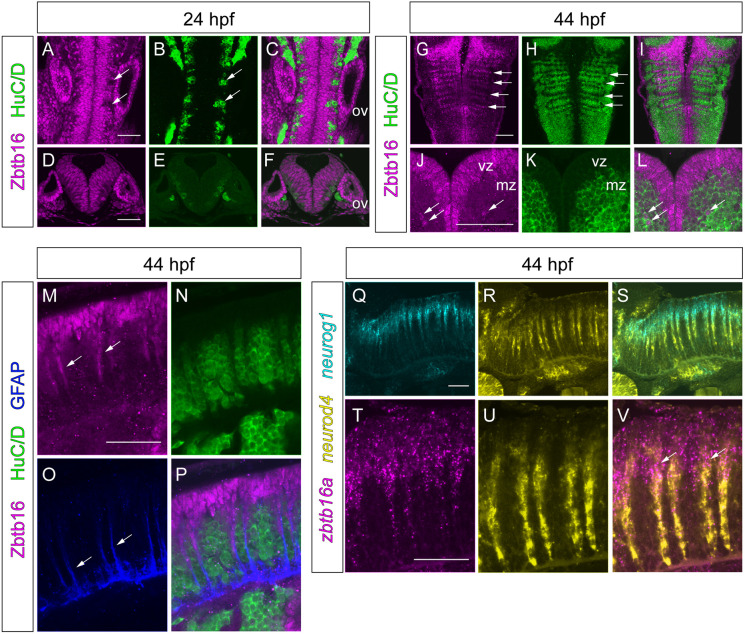
Expression of Zbtb16 during neurogenesis. (A-L) Immunofluorescence for Zbtb16 and HuC/D at 24 and 44 hpf. Dorsal views (A-C,G-I) and transverse sections (D-F,J-L) of the hindbrain. G-I are coronal slices at the level of the mantle zone. Arrows indicate postmitotic neurons (A,B), neurogenic zones (G,H) or Zbtb16+HuC/D double-positive cells (J,L). (M-P) Immunofluorescence for Zbtb16, HuC/D and GFAP at 44 hpf. Side views of the hindbrain, anterior to the left. Arrows indicate Zbtb16-positive migrating progenitors (M) or GFAP-positive glial fibres (O). (Q-V) Two-colour fluorescent ISH for neurog1 and neurod4 (Q-S) or zbtb16a and neurod4 (T-V) at 44 hpf. Side views of the hindbrain, anterior to the left. Arrows indicate overlap of zbtb16a and neurod4 expression. All images are slices from confocal z-stacks. mz, mantle zone; ov, otic vesicle; vz, ventricular zone. Scale bars: 50 µm.
Acknowledgments
This image is the copyrighted work of the attributed author or publisher, and
ZFIN has permission only to display this image to its users.
Additional permissions should be obtained from the applicable author or publisher of the image.
Full text @ Development
Your Input Welcome
Thank you for submitting comments. Your input has been emailed to ZFIN curators who may contact you if
additional information is required.
Oops. Something went wrong. Please try again later.