Image
Figure Caption
Figure 6
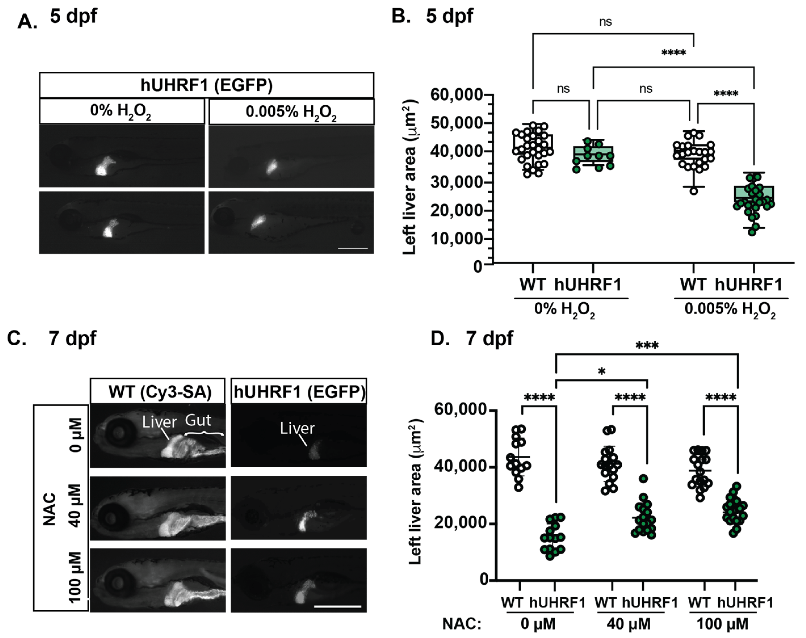
Figure 6. The small liver phenotype caused by hUHRF1 overexpression is enhanced by H2O2 exposure and reversed by NAC treatment. (A) hUHRF1 transgenic larvae and their non-transgenic siblings were untreated or treated with 0.005% H2O2 for 40–42 h and collected at 120 hpf. The left liver area was measured using the EGFP fluorescence (hUHRF1 transgenics) or CY3-SA staining (WT). (B) Left liver lobe area is significantly smaller in hUHRF1 larvae treated with 0.005% H2O2 compared to non-transgenic siblings treated with H2O2 and compared to untreated hUHRF1 larvae. The median is indicated by the horizontal line in the box and the whiskers represent the range of measurements. (C) hUHRF1 transgenic larvae and their non-transgenic siblings were untreated or treated with 40 or 100 μM of NAC from 4 dpf to 7 dpf and imaged at 7 dpf for left liver area on EGFP fluorescence (transgenics) or CY3-SA staining (controls). (D) Liver size is significantly smaller in hUHRF1 larvae compared to non-transgenic siblings at 7 dpf and NAC treatment increases the size of the liver in transgenic larvae. Experiments were performed in 2–3 clutches with at least 9 animals per clutch. Each dot represents 1 liver. Scale bar: 500 µm. Values are expressed as the mean ± SD and were compared by two-way ANOVA with Tukey’s multiple comparisons test (B,D). Significant differences are indicated as follows: ns p > 0.05 (non-significant), * p < 0.05, *** p < 0.001, **** p < 0.0001.
Acknowledgments
This image is the copyrighted work of the attributed author or publisher, and
ZFIN has permission only to display this image to its users.
Additional permissions should be obtained from the applicable author or publisher of the image.
Full text @ Cancers

