Fig. 2
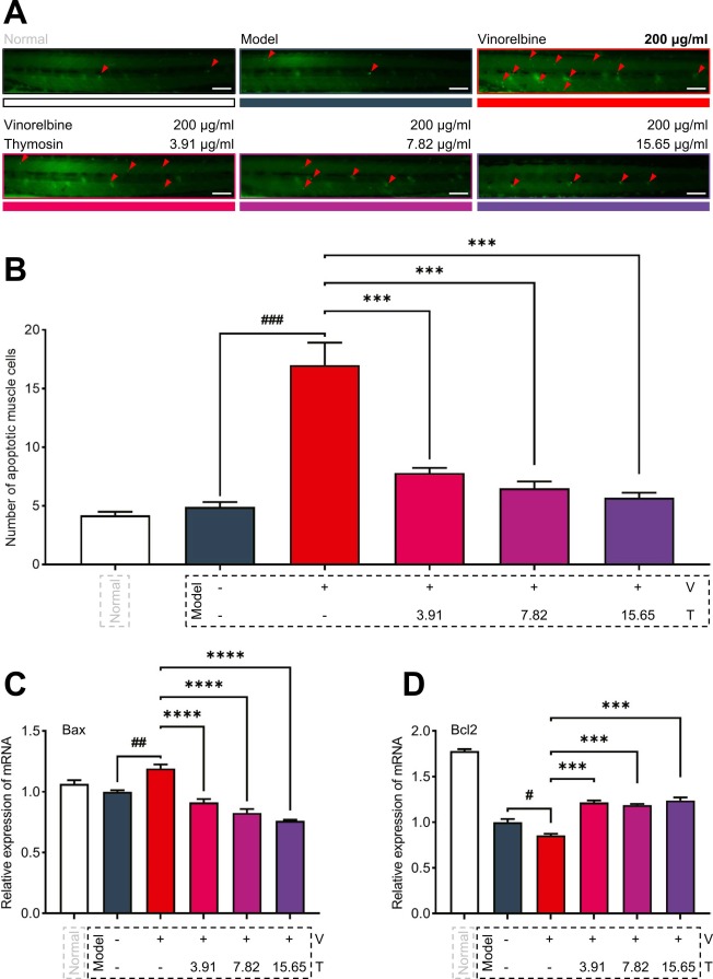
Fig. 2. Thymosin alleviates vinorelbine-induced apoptosis of muscle cells in the xenotransplanted zebrafish model. (A) Representative images of xenotransplanted zebrafish in different groups. The acridine orange, a nucleic acid intercalating dye that emits green fluorescence when bound to dsDNA and selectively stains apoptotic cells, is utilized here. Red arrows indicate apoptotic muscle cells. Scale bar, 50 µm. (B) The number of apoptotic muscle cells after treatment with vinorelbine and thymosin for 18 h. (C) The transcription levels of apoptosis-promoting factor, Bax, and (D) apoptosis inhibitor, Bcl2, after treatment of vinorelbine and thymosin for 18 h. The mRNA level of the model control group was set at 1 in the correlation analysis of qRT-PCR results. Data are expressed as mean ± SEM. n = 10. ∗∗∗P < 0.001, ∗∗∗∗P < 0.0001 compared to the vinorelbine treatment group; #P < 0.05, ##P < 0.01, ###P < 0.001, comparison between the model control group and the vinorelbine treatment group. Abbreviations: T: thymosin; V: vinorelbine.

