
IMAGE
Fig. 4
- ID
- ZDB-IMAGE-230912-10
- Publication
- Zhang et al., 2023 - Knockout of miR-184 in zebrafish leads to ocular abnormalities by elevating p21 levels
- All Figures
- Figures for Zhang et al., 2023
Image
|
Figure Caption
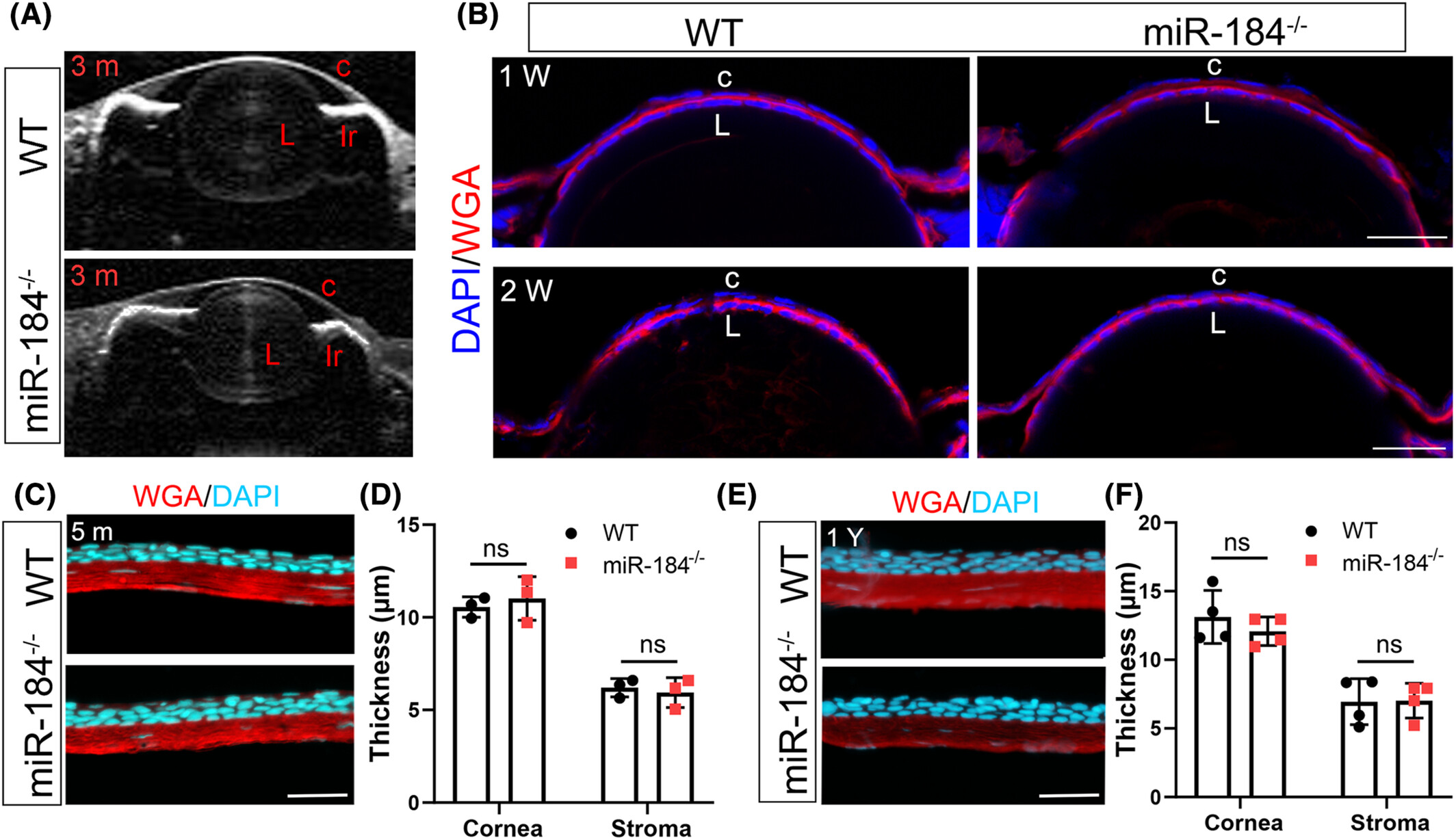
Fig. 4
No corneal abnormity was observed in miR-184−/− zebrafish. (A) The anterior corneal morphology of 3-month-old miR-184−/− zebrafish was observed with a AS-OCT. L, lens; C, cornea; Ir, iris. (B) The cornea development was detected in embryonic WT and miR-184−/− zebrafish. The corneal epithelia were labeled with DAPI staining, while the stroma was stained with Alexa Fluor™ 594-conjugated WGA. L, lens; C, cornea. Bar, 25 μm. (C and D) The corneal structure (C) and thickness (D) were detected in 5-month-old WT and miR-184−/− zebrafish. Student t-test was used for statistical analysis, n = 3. ns, no significant. Bar, 50 μm. (E and F) The corneal structure (E) and thickness (F) were detected in 1-year-old WT and miR-184−/− zebrafish. Student t-test was used for statistical analysis, n = 4. ns, no significant. Bar, 50 μm.
Acknowledgments
This image is the copyrighted work of the attributed author or publisher, and
ZFIN has permission only to display this image to its users.
Additional permissions should be obtained from the applicable author or publisher of the image.
Full text @ FASEB J.