
Fig. 3
- ID
- ZDB-IMAGE-230904-3
- Publication
- Bi et al., 2023 - Polysarcosine-based lipid formulations for intracranial delivery of mRNA
- All Figures
- Figures for Bi et al., 2023
|
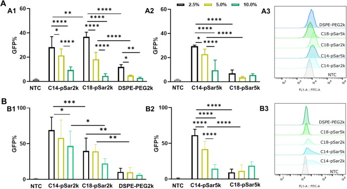
Fig. 3 Lipoplexes containing GFP mRNA and PE-LR labeled liposomes were incubated with (A) Jurkat T cells and (B) Hela cells for 24 h to examine transfection efficiency. Lipoplexes were prepared by incubating equal volume of mRNA and liposomes at N/P 10. At the end of incubation, cells were harvested and quantified by flow cytometry, as demonstrated in (A1, A2) of Jurkat T cells and (B1, B2) Hela cells. Representative images of (A3) Jurkat T cells and (B3) Hela cells, after transected with mRNA lipoplexes composed of 2.5% DSPE-PEG2k or pSar-lipid-polyplexes. All the data was averaged from three independent experiments (n = 9).