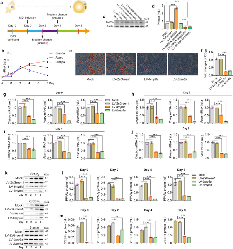
IMAGE

Fig. 3

ID
ZDB-IMAGE-230814-264
Source
Figures for Zhong et al., 2023
Image
Figure Caption

Fig. 3 Stably overexpressing zebrafish bmp8a or mouse Bmp8a inhibits adipogenesis.

a Protocol for effective differentiation of 3T3-L1 cells into adipocytes. b The mRNA expression pattern of mouse Bmp8a, Pparγ and C/ebpα during 3T3-L1 cells differentiated into adipocytes (n = 3). c, d Immunoblot analysis of mouse BMP8A protein expression in 3T3-L1 cells (Mock), stably overexpressed empty plasmid in 3T3-L1 cells (LV-ZsGreen1), stably overexpressed mouse Bmp8a in 3T3-L1 cells (LV-Bmp8a), 3T3-L1 cells infected with scramble shRNA lentivirus (LV-shRNA-scrambled), and knockdown mouse Bmp8a in 3T3-L1 cells (shRNA-Bmp8a#1 and shRNA-Bmp8a#2). BMP8A protein expression levels were quantified by ImageJ software and normalized to the amount of β-actin (d,n = 3). e, f After induction of adipogenic differentiation, differentiated 3T3-L1 adipocytes (Mock, LV-ZsGreen1, LV-bmp8a, and LV-Bmp8a) were stained with Oil Red O and subjected to OD492 quantifications (n = 3). Scale bar = 20 µm. gj On the day after induction as indicated, expressions of adipogenic genes (Cebpα, Pparγ, and Fasn) were examined at the mRNA level by qPCR (n = 3). km On the day after induction, as indicated, the protein levels of PPARγ and C/EBPα detected by Immunoblot. Protein expression levels were quantified using ImageJ software and normalized to the amount of β-actin (l, m, n = 3). Data were representative of at least three independent experiments. Data were analyzed by One-way ANOVA and presented as mean ± SD (**p < 0.01, ***p < 0.001).

Acknowledgments
This image is the copyrighted work of the attributed author or publisher, and ZFIN has permission only to display this image to its users. Additional permissions should be obtained from the applicable author or publisher of the image. Full text @ Commun Biol