
Fig. 1
- ID
- ZDB-IMAGE-230810-1
- Genes
- Publication
- Yao et al., 2023 - DExH/D RNA helicase 15 regulates zebrafish intestinal development through the Wnt signaling pathway
- All Figures
- Figures for Yao et al., 2023
|
Fig. 1
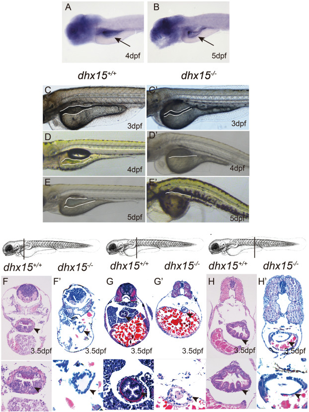
Fig. 1. Dhx15 knockout exhibits early intestinal structure abnormalities in zebrafish. (A-B) WISH analysis of RNA probe dhx15 in zebrafish at 4 dpf (A) and 5 dpf (B). The data showed dhx15 expressed in the intestine of zebrafish at 4 dpf and 5 dpf. (C-E C′-E') Lateral view of zebrafish embryos. Bright field images of dhx15+/+ and dhx15−/− zebrafish under microscope (Leica M50, ×10). The foregut enlargement of dhx15−/− embryos was not obvious compared with that in the widetype (the white line). (F-H F′-H′) H & E staining of the sagittal section from dhx15+/+ and dhx15−/− zebrafish at 3.5 dpf. The figures showed that the intestinal epithelium of dhx15−/− zebrafish (F′-H′ × 20, × 63) embryos was lacked folds, and had thin walls compared with the widetype (F H × 20, × 63). The position of the section was shown above the H & E staining images. The arrow and white line indicate the intestine of zebrafish. The arrow heads indicate the epithelium of the intestine.