IMAGE

Fig. 2.

ID
ZDB-IMAGE-230801-10
Source
Figures for Jubber et al., 2023
Image
Figure Caption

Fig. 2.

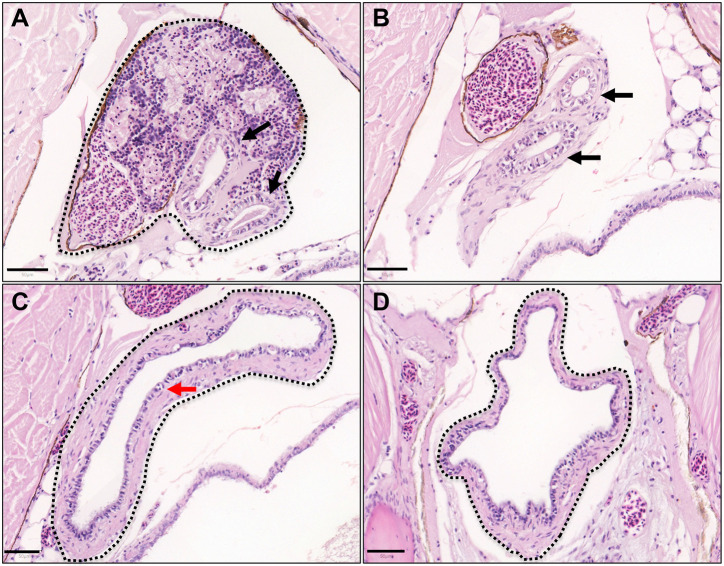
Anatomy of adult zebrafish mesonephric ducts and urinary bladder. Haematoxylin and Eosin stained adult zebrafish sections in transverse orientation from cranial to caudal. (A) Kidney (black outline) converges onto two mesonephric ducts (black arrows). (B) Two mesonephric ducts (black arrows) distal to the kidney in the retroperitoneum. (C) The two mesonephric ducts unite into a single urinary bladder lined with epithelial cells (red arrow). (D) Inferior aspect of the urinary bladder. Scale bars: 50 μm.

Acknowledgments
This image is the copyrighted work of the attributed author or publisher, and ZFIN has permission only to display this image to its users. Additional permissions should be obtained from the applicable author or publisher of the image. Full text @ Dis. Model. Mech.