
|
Fig. 6
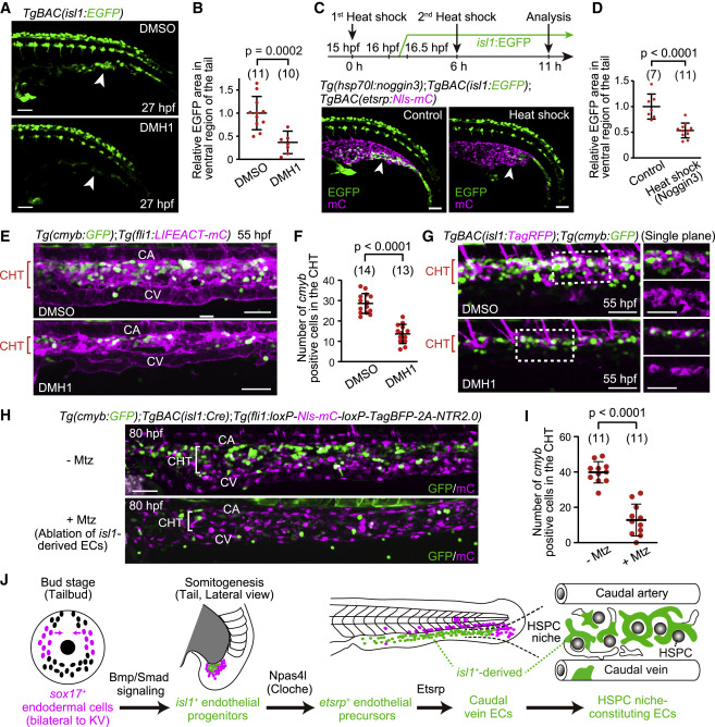
Figure 6. isl1+ endothelial progenitors are specified by Bmp-Smad signaling and constitute the vascular HSPC niche (A) TgBAC(isl1:EGFP) embryos treated with DMSO or DMH1 at 15 hpf (i.e., in advance of isl1:EGFP expression [16–16.5 hpf]) and observed 12 h after the treatment. The number of isl1:EGFP+ cells in the tail is markedly decreased by DMH1 treatment (arrowheads). (B) Quantitative analysis of the data shown in (A). isl1:EGFP area in the ventral region of the tail in embryos treated with DMH1 relative to that in embryos treated with DMSO. EGFP area is measured as in Figure S5B. Data are mean ± SD. (C) Tg(hsp70l:noggin3);TgBAC(isl1:EGFP);TgBAC(etsrp:Nls-mCherry) embryos without (control) or subjected to heat shock twice (upper, schematic illustration). The number of isl1:EGFP+ cells in the tail is markedly decreased by Noggin3 overexpression (arrowheads). (D) Quantitative analysis of the data shown in (C). isl1:EGFP area as in (B) in the heat-shocked embryos relative to that in embryos without heat shock. Data are mean ± SD. (E) Tail of Tg(cmyb:GFP);Tg(fli1:LIFEACT-mCherry) embryos (55 hpf) treated with DMSO or DMH1 for 12 h starting at 15 hpf. (F) Number of cmyb:GFP+ cells in the CHT regions of DMSO- or DMH1-treated embryos (54–55 hpf), as observed in (E). Data are mean ± SD. (G) Tail of TgBAC(isl1:TagRFP);Tg(cmyb:GFP) embryos (55 hpf) treated with DMSO or DMH1 for 12 h starting at 15 hpf as in (E). Left, projection view; right, single planes of the boxed areas. (H) Tail of TgBAC(isl1:Cre);Tg(fli1:loxP-Nls-mCherry-loxP-TagBFP-2A-NTR2.0) larvae (80 hpf) treated with or without Mtz for 24 h starting at 35 hpf. (I) Number of cmyb:GFP+ cells in the CHT regions of larvae (80–81 hpf) treated with or without Mtz, as observed in (H). Data are mean ± SD. (J) Schematic representation of the development of caudal vein ECs from sox17+ endodermal cells. Scale bars, 50 μm. See also Figure S5.
Reprinted from Developmental Cell, 58(3), Nakajima, H., Ishikawa, H., Yamamoto, T., Chiba, A., Fukui, H., Sako, K., Fukumoto, M., Mattonet, K., Kwon, H.B., Hui, S.P., Dobreva, G.D., Kikuchi, K., Helker, C.S.M., Stainier, D.Y.R., Mochizuki, N., Endoderm-derived islet1-expressing cells differentiate into endothelial cells to function as the vascular HSPC niche in zebrafish, 224-238.e7, Copyright (2023) with permission from Elsevier. Full text @ Dev. Cell