
|
Fig. 4
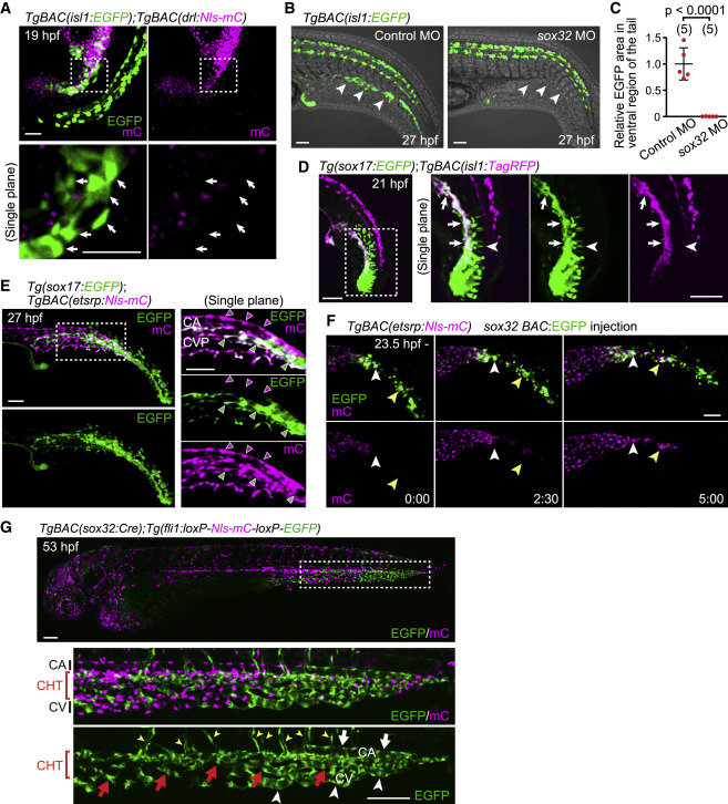
Figure 4. The isl1+ endothelial progenitors do not originate from the lateral plate mesoderm but from cells expressing sox32 and sox17 (A) TgBAC(isl1:EGFP);TgBAC(drl:Nls-mCherry) embryo (19 hpf). Upper, projection view; lower, single confocal plane of the boxed area. isl1:EGFP+ cells (arrows) do not express drl:mCherry. (B) TgBAC(isl1:EGFP) embryos (27 hpf) injected with control MO or sox32 MO. Ventral isl1:EGFP+ cells are not detected in sox32 morphants (arrowheads). (C) Quantitative analyses of the data shown in (B). EGFP area in the ventral region of the tail in embryos injected with sox32 MO relative to that in embryos injected with control MO as shown in Figure S5B. Data are mean ± SD. (D) Tg(sox17:EGFP);TgBAC(isl1:TagRFP) embryo (21 hpf). Left, projection view; right, single confocal plane of the boxed area. Arrows point to sox17:EGFP/isl1:TagRFP double-positive cells. Arrowheads point to sox17:EGFP single-positive notochord cells. (E) Tg(sox17:EGFP);TgBAC(etsrp:Nls-mCherry) embryo (27 hpf). Left, projection view; right, single plane of the boxed area. sox17:EGFP expression in the CVP (green arrowheads) but not in the CA (magenta arrowheads). (F) Time-sequential images in the tail of TgBAC(etsrp:Nls-mCherry) (from 23.5 hpf) injected with sox32 BAC:EGFP plasmid that drives EGFP expression under the control of a sox32 BAC promoter in a mosaic manner. sox32:EGFP+ cells differentiate into etsrp:mCherry+ endothelial precursors (arrowheads point to individual cells). (G) TgBAC(sox32:Cre);Tg(fli1:loxP-Nls-mCherry-loxP-EGFP) embryo (53 hpf). The boxed area is enlarged (middle and bottom). Cre-driven EGFP expression in ECs is restricted to the tail, especially in the CHT (red arrows) and CHT-derived vISVs (yellow arrowheads). In the most posterior end, it is found in almost all ECs including the CA (white arrowhead), CHT (red arrows), and CV (white arrows). Scale bars: 50 μm in (A), (B), and (D)–(F) and 100 μm in (G). See also Figure S4.
Reprinted from Developmental Cell, 58(3), Nakajima, H., Ishikawa, H., Yamamoto, T., Chiba, A., Fukui, H., Sako, K., Fukumoto, M., Mattonet, K., Kwon, H.B., Hui, S.P., Dobreva, G.D., Kikuchi, K., Helker, C.S.M., Stainier, D.Y.R., Mochizuki, N., Endoderm-derived islet1-expressing cells differentiate into endothelial cells to function as the vascular HSPC niche in zebrafish, 224-238.e7, Copyright (2023) with permission from Elsevier. Full text @ Dev. Cell