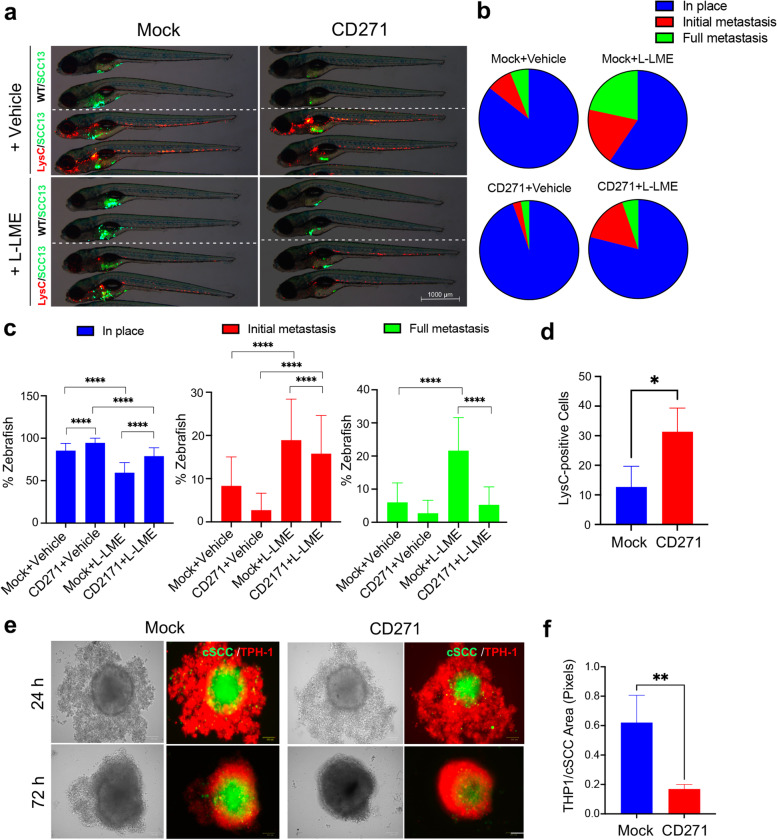
CD271 expression increases the immune cell recruitment in vivo and in vitro a Representative images of mock and CD271-overexpressing SCC13 cells (green) injected into WT (indicated as WT/SCC13 in the figure) or Tg(lysC:DcRed)nz50 (indicated as LysC/SCC13) zebrafish with or without the simultaneous treatment with the L-LME. In the Tg(lysC:DcRed2)nz50 zebrafish, leucocytes possess a red fluorescence. Scale bar = 500 µm. b and c cSCC metastases quantified and classified as Full metastases, Initial metastases, or In place. d Quantification of LysC-positive cells in zebrafish injected with mock or CD271-overexpressing cells e SCC13 cells were stained with Vybrant DIO Cell-Labeling Solution (green) and seeded in a 96-well plate coated with 1,5% of agar. cSCC spheroids were transduced by infection with mock or CD271 viral vectors. After 72 h, THP-1 cells were stained with Vybrant DII Cell-Labeling Solution (red) and co-cultured with cSCC spheroids. Pictures were made after 24 and 72 h from the THP-1 seeding. Scale bar = 100µm. f The invasion area of THP-1 was measured by ImageJ software. Results are shown as the mean percentage of three independent experiments (minimum of 20 injected zebrafish/condition). Statistical analysis was performed using the two-way ANOVA and multiparametric T-test. **0.001 < p < 0.01, ****p < 0.0001
Acknowledgments
This image is the copyrighted work of the attributed author or publisher, and
ZFIN has permission only to display this image to its users.
Additional permissions should be obtained from the applicable author or publisher of the image.
Full text @ J. Exp. Clin. Cancer Res.
Your Input Welcome
Thank you for submitting comments. Your input has been emailed to ZFIN curators who may contact you if
additional information is required.
Oops. Something went wrong. Please try again later.