
|
Fig. 2
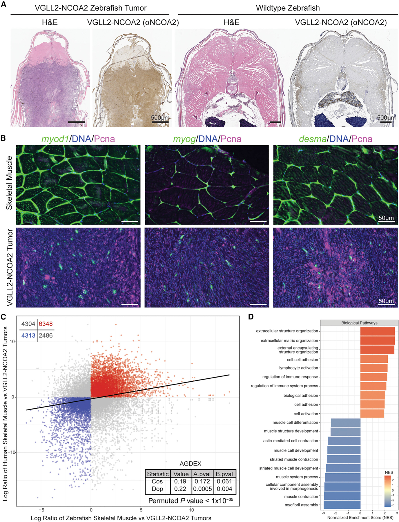
(A) Transverse sections through the GFP+ tumors were stained with H&E, and the subsequent step section stained with a human anti-NCOA2 antibody to detect the VGLL2-NCOA2 fusion. All n = 9 zebrafish tumors stained were positive. A transverse section was taken through a control wild-type zebrafish, and shown are the H&E and subsequent anti-NCOA2 antibody stain. Scale bars, 500 µm.
(B) Representative RNAscope of transverse sections from wild-type zebrafish (portraying normal skeletal muscle expression) or zebrafish with VGLL2-NCOA2 tumors showing expression of myod1, myog, or desma (green), Pcna (purple), or DNA (blue). Scale bars, 50 µm. Five wild-type or tumor samples were assessed in total, with results presented in Table S3.
(C) RNA-seq data of zebrafish VGLL2-NCOA2 tumors (n = 18) and mature skeletal muscle (n = 7) were compared with their human counterpart (VGLL2-NCOA2 tumors n = 5 from Watson et al.4 and mature skeletal muscle n = 803 from GTEx version 8) in an AGDEX analysis. Plotted in blue and red are genes that are shared as differentially expressed between the zebrafish and human VGLL2-NCOA2 tumors. The statistics of the AGDEX analysis are shown in the bottom right, as well as the p value generated by permutation analysis of randomly sampled data.
(D) Gene set enrichment analysis associated with the n = 6,348 genes upregulated or n = 4,313 downregulated in (C). Plotted is the normalized enrichment score for each sub-ontology.