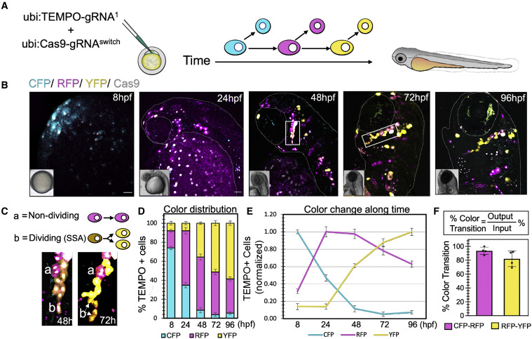
Fig. 2 TEMPO enables sequential cell labeling in live zebrafish embryos
(A) Co-injection of Tol1 integrative ubiquitous TEMPO and Cas9 constructs into 1-cell-stage zebrafish embryos. TEMPO transitions analyzed in live embryos between 8 and 96 h post fertilization (hpf).
(B) Snapshots of ubiquitously expressed TEMPO reporter cascade in live zebrafish embryos. Scale bars, 50 μm.
(C) Insets highlight a cell clone imaged at 48 and 72 h including (a) non-dividing cells which do not transition in the cascade and (b) dividing cells which transition to the last color (YFP).
(D) Changes in TEMPO color distribution (percentage of fluorescent cells for each color reporter along time) demonstrate efficient reporter transitions in a predefined order in developing zebrafish (8 h, n = 8; 24 h, n = 5; 48 h, n = 6; 72 h, n = 4; 96 h, n = 4, error bars represent SEM).
(E) Normalization of data shown in (D) to the maximum fluorescence for each reporter along time.
(F) Efficiency of color transition, defined as the ratio between the final proportion of TEMPO+ cells at 96 h (output) over the proportion of initial TEMPO+ cells at 8 h (input) for any given reporter transition. Data are shown as percentage ± SEM.

