IMAGE

Figure 9

ID
ZDB-IMAGE-230707-119
Source
Figures for Van Dyck et al., 2023
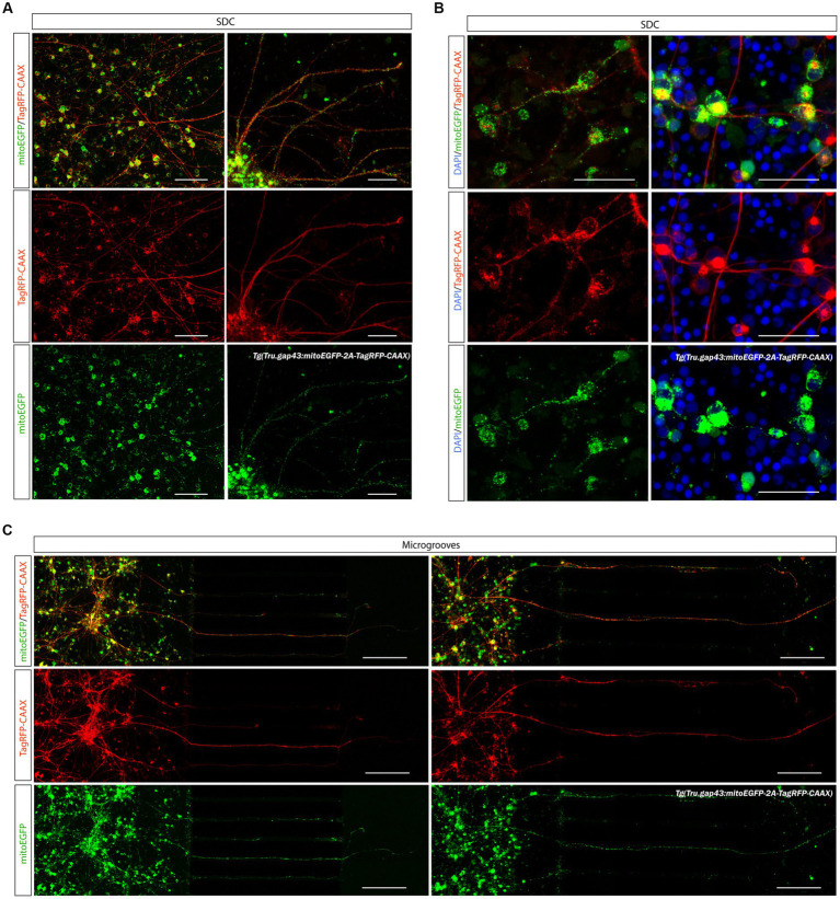
Image
Figure Caption

Figure 9

Visualization of mitochondria in adult zebrafish RGCs in microfluidic cultures. The recently developed Tg(Tru.gap43:mitoEGFP-2A-tagRFP-CAAX)ulb17 (gapmito) reporter line enables to study mitochondrial motility in outgrowing and regenerating RGCs. (A) Confocal live cell images of gapmito retinal neurons at DIV 3 in an open compartment (SOC450) MFD indicate that outgrowing RGCs express RFP in membranes (middle panels) and EGFP in mitochondria in axons, dendrites and somata (bottom panels). (B) Detailed live cell images (left panels), as well as pictures of end-point DAPI-stained cultures of isolated neurons (right panels) in the SDC illustrate that mitochondria in the different neuronal compartments can be visualized with subcellular resolution. (C) Likewise in the microgrooves and AC, individual mitochondria can be identified in outgrowing gapmito RGC axons at DIV 3. These results demonstrate how the usefulness of this novel reporter line to characterize mitochondrial dynamics during axonal outgrowth or regeneration. Scale bars: 100 μm (A,C), 50 μm (B). AC, axonal compartment; DIV, days in vitro; MFD, microfluidic device; RGC, retinal ganglion cell; SDC, somatodendrtic compartment.

Acknowledgments
This image is the copyrighted work of the attributed author or publisher, and ZFIN has permission only to display this image to its users. Additional permissions should be obtained from the applicable author or publisher of the image. Full text @ Front. Mol. Neurosci.