
Figure 5
- ID
- ZDB-IMAGE-230707-115
- Publication
- Van Dyck et al., 2023 - A new microfluidic model to study dendritic remodeling and mitochondrial dynamics during axonal regeneration of adult zebrafish retinal neurons
- All Figures
- Figures for Van Dyck et al., 2023
|
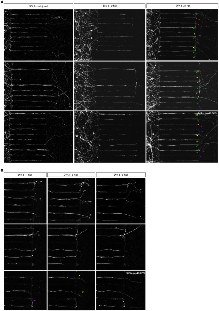
Figure 5
Axotomy and regeneration of adult zebrafish RGCs at DIV 3–4. Representative confocal live cell microscopy images taken before, upon and after injury in the same section of the AC, disclose that adult zebrafish RGCs regenerate spontaneously upon