
Figure 5
- ID
- ZDB-IMAGE-230630-49
- Publication
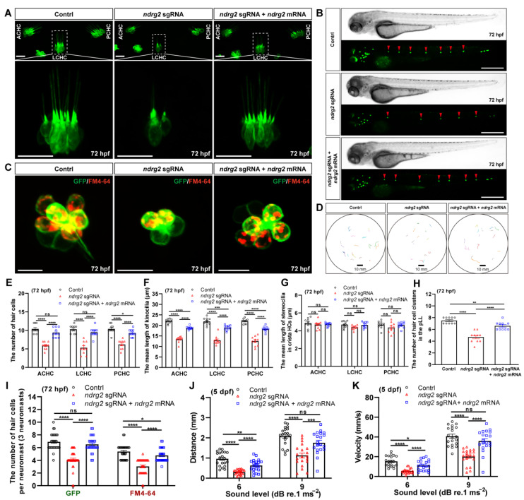
- Wang et al., 2023 - The ndrg2 Gene Regulates Hair Cell Morphogenesis and Auditory Function during Zebrafish Development
- All Figures
- Figures for Wang et al., 2023
|
Figure 5
Knockout of the