
Fig. 8
- ID
- ZDB-IMAGE-230627-8
- Publication
- Flex et al., 2022 - Dominantly acting KIF5B variants with pleiotropic cellular consequences cause variable clinical phenotypes
- All Figures
- Figures for Flex et al., 2022
|
Fig. 8
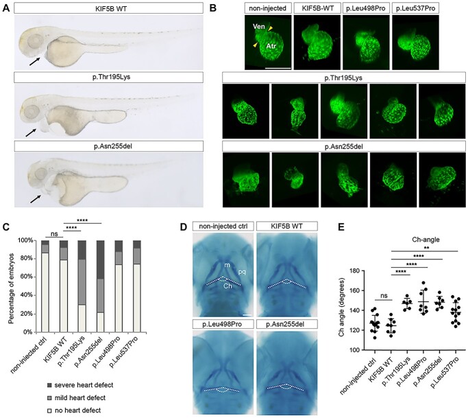
KIF5B is important for proper heart formation and contributes to craniofacial development in zebrafish. (A) Representative images of KIF5B variant-injected embryos for the different morphological classes of heart defects (from top to bottom, no defects, mild and severe defects). (B) Confocal imaging of Tg(cmlc2:eGFP) zebrafish transgenic hearts at 2 days post-fertilization (dpf). At this stage the zebrafish heart is formed by two chambers, the ventricule (Ven) and the atrium (Atr) separated by a contractile ring (yellow arrowheads). Injected transgenic embryos with the different KIF5B variants at 1-cell stage were imaged at 2 dpf, revealing heart formation defects specifically in embryos expressing KIF5BAsn255del variants. Scale bars: 150 μm. (C) Heart defects quantification of 2 dpf larvae injected with KIF5B alleles. With respect to non-injected controls and wild type KIF5B-injected embryos, larvae injected with the KIF5BAsn255del and the KIF5Bp.Thr195Lys variants show a significant increase in the number of embryos with mild and severe heart defects. (****) P-value < 0.0001. Ven: ventricle, Atr: atrium. (D) Alcian blue staining at 3 dpf of zebrafish larvae injected at 1-cell stage with (top. right) wild-type KIF5B (bottom, left) KIF5BLeu495Pro (bottom, right) KIF5BAsn255del, compared to the non-injected larvae (top, left). White dashed lines label the ceratohyal cartilages. (E) The ceratohyal angle (Ch-angle) for each condition was measured, revealing a wider angle at the ceratohyal cartilage intersection for all injected variants compared to non-injected control larvae or wild-type KIF5B. ch: ceratohyal; m: Meckel’s; pq: palatoquadrate. (**) P-value = 0.0081 and (****) < 0.0001. Scale bars: 100 μm.