
Fig. 4
- ID
- ZDB-IMAGE-230627-4
- Publication
- Flex et al., 2022 - Dominantly acting KIF5B variants with pleiotropic cellular consequences cause variable clinical phenotypes
- All Figures
- Figures for Flex et al., 2022
|
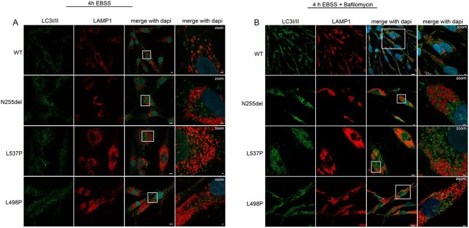
Fig. 4
Disease-causing KIF5B mutations affect autophagosomes morphology and subcellular localization. (A) Confocal microscopy analyses show a peculiar subcellular localization of autophagosomes in patients’ cells compared to control cells. In particular, these organelles localize near the nucleus and are enlarged compared to that observed in control cells, and a few number colocalized with lysosomes residing in the peripheral region of the cell. Scale bars are respectively 10 μm (left) and 2 μm (right). (B) In these panels, this behavior is exacerbated by treatment with bafilomycin. Scale bars are respectively 10 μm (left) and 5 μm (right). The cells were stained with antibodies against Lamp1 (lysosomes marker, red), LC3I/II (autophagosomes marker, green) and DAPI (DNA marker, blue).