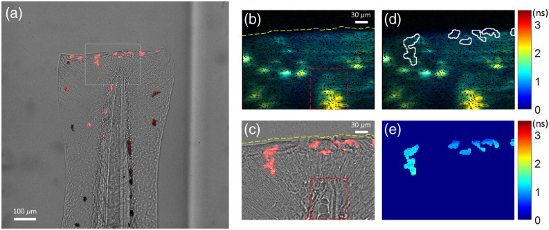
Light-sheet imaging of mCherry intensity and NAD(P)H lifetimes in live neutrophils in vivo in a zebrafish caudal fin wound model. (a) The intensity channel of the light-sheet FLIM system shows the superimposed brightfield image of 3 dpf wounded zebrafish tail and light-sheet-excited fluorescence intensity of mCherry+ neutrophils at the wound site (Video 2, MPEG, 2.1 MB [URL: https://doi.org/10.1117/1.JBO.28.6.066502.s2]). The white rectangle shows the corresponding field of view of the smaller SPAD array sensor. (b) NAD(P)H mean fluorescence lifetime (τm) image of the tail (from the SPAD array with 10 s integration). The transection wound (yellow dashed line) and the tip of the notochord (red dashed box) are visible in the lifetime image. (c) Corresponding field of view from the cropped sCMOS image shows mCherry+ neutrophils recruited to the wound site. (d) Pixel mask of neutrophil cells from the mCherry channel applied to the NAD(P)H lifetime image. (e) Masked NAD(P)H lifetime map of the neutrophils.
Acknowledgments
This image is the copyrighted work of the attributed author or publisher, and
ZFIN has permission only to display this image to its users.
Additional permissions should be obtained from the applicable author or publisher of the image.
Full text @ J. Biomed. Opt.
Your Input Welcome
Thank you for submitting comments. Your input has been emailed to ZFIN curators who may contact you if
additional information is required.
Oops. Something went wrong. Please try again later.