
|
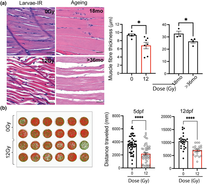
FIGURE 2
Zebrafish larvae showed reduced muscle fibre thickness and mobility following irradiation. (a) Representative photomicrographs of haematoxylin and eosin (H&E) staining of zebrafish muscle from either 12 dpf larvae with and without irradiation (top left panel), and middle aged and geriatric adults (bottom left panel) (scale 25 μm) and quantification of muscle fibre thickness in larvae following irradiation and in middle aged (18 months) and geriatric zebrafish (>36 months) (right panels). Data shown as mean ± SEM. Each dot represents an animal. (b) Representative example of distance travelled by zebrafish over 30 min in a 24‐well plate at 5 (