
Image
|
Figure Caption
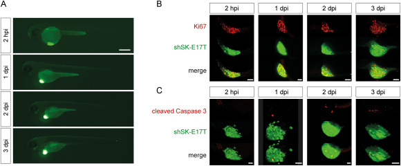
Fig. 1 Fig. 1. shSK-E17T Ewing sarcoma zebrafish xenografts A) Zebrafish xenografted with GFP-expressing shSK-E17T Ewing sarcoma cells (green) imaged on a fluorescence microscope over consecutive days from 2 hpi to 3 dpi. B) Immunostaining for human Ki67 revealing proliferating tumor cells and C) for cleaved Caspase 3 demarcating apoptotic cells at different time points from 2 hpi to 3 dpi. (B, n: 2 hpi = 5, 1 dpi = 7, 2 dpi = 5, 3 dpi = 6); (C, n: 2 hpi = 6, 1 dpi = 8, 2 dpi = 5, 3 dpi = 7). Scale bars are 250 μm in A and 50 μm in B and C.
Acknowledgments
This image is the copyrighted work of the attributed author or publisher, and
ZFIN has permission only to display this image to its users.
Additional permissions should be obtained from the applicable author or publisher of the image.
Full text @ Cancer Lett.