Fig. 1
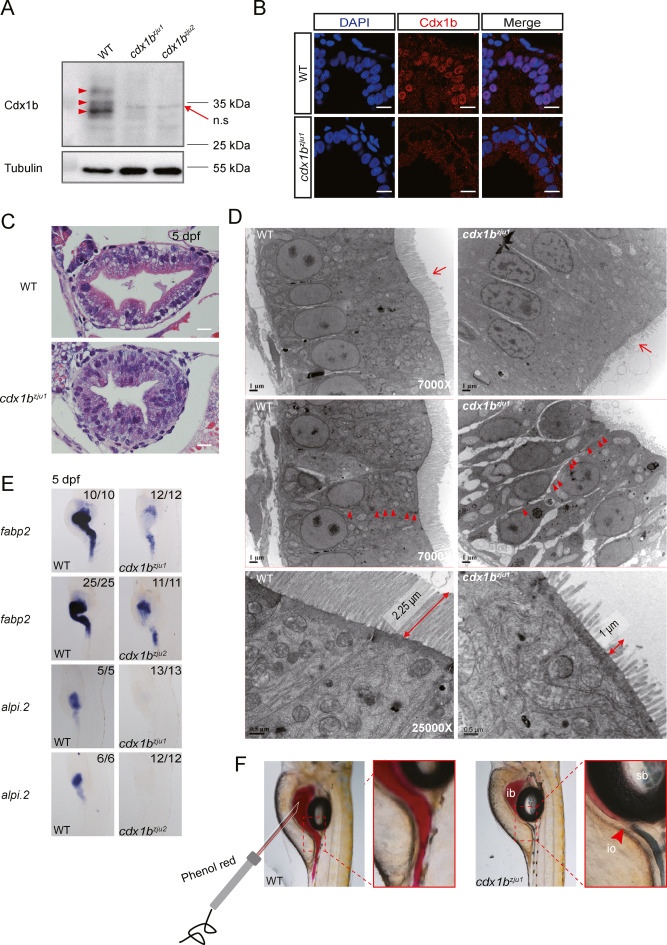
Fig. 1. Cdx1b is essential for the development of a functional intestine in zebrafish. A: Western blot showing the depletion of Cdx1b protein in two cdx1b mutants cdx1bzju1/zju1 (cdx1bzju1) and cdx1bzju2/zju2 (cdx1bzju2) compared with WT at 5 dpf. In WT, Cdx1b showed three isoforms (red arrow head). B: Immunostaining of Cdx1b (red) in the intestines of WT and cdx1bzju1/zju1. Cdx1b is a nuclear localized protein. DAPI stains the nuclei. Scale bar, 10 μm. C: HE staining showing the disorganized gut epithelium in cdx1bzju1/zju1 compared with WT at 5 dpf. Scale bar, 10 μm. D: TEM images showing the shortened microvilli and loosened cell-cell junction in cdx1bzju1/zju1 compared with WT at 5 dpf. Red arrow, microvilli; double headed arrow, length of microvilli; arrow head, cell-cell junction. Scale bar, 1 μm or 0.5 μm (bottom two images). E: WISH showing the decreased expression of fabp2 and near absence of alpi.2 in the cdx1bzju1/zju1 and cdx1bzju2/zju2 intestine compared with that in WT at 5 dpf. F: Images showing the blockage of intestinal fluid by visualizing the injected phenol-red at the junction between the intestinal bulb (ib) and mid-intestine in cdx1bzju1/zju1 compared with WT. n.s, non-specific band. io, intestinal obstruction; sb, swim bladder.
Reprinted from Journal of genetics and genomics = Yi chuan xue bao, 49(12), Jin, Q., Gao, Y., Shuai, S., Chen, Y., Wang, K., Chen, J., Peng, J., Gao, C., Cdx1b protects intestinal cell fate by repressing signaling networks for liver specification, 1101-1113, Copyright (2022) with permission from Elsevier. Full text @ J. Genet. Genomics

