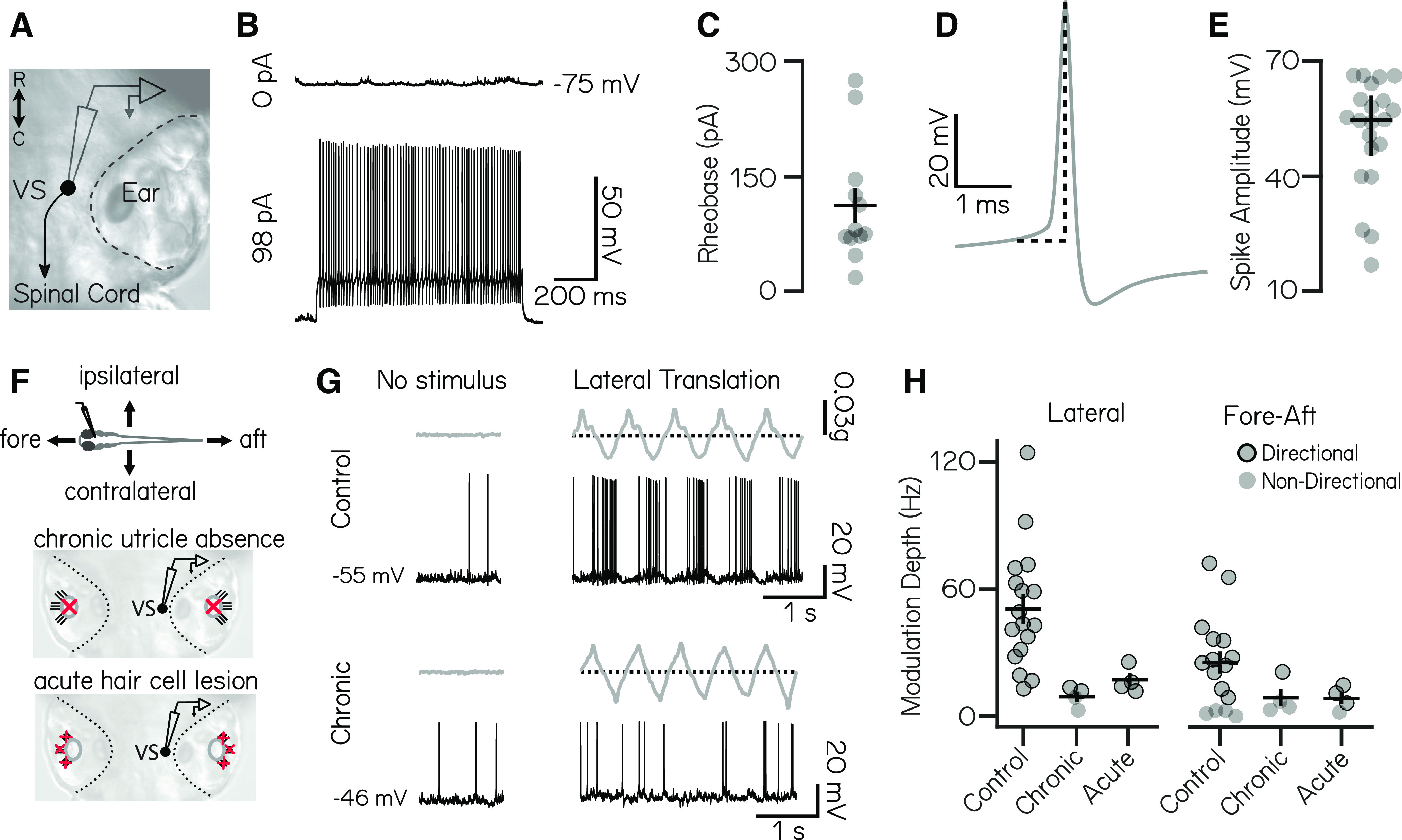
Vestibulospinal neurons encode utricle-derived body translation. A, Schematic of spinal-projecting vestibulospinal (VS) neuron targeted for electrophysiology. B, Vestibulospinal membrane potential at rest (top, 0 pA) and in response to current injection (bottom, 98 pA). C, Rheobase (mean ± SEM) across 12 nonspontaneously active cells. D, Example action potential waveform with amplitude (dotted line). E, Action potential average amplitude (median ± interquartile range) across 21 cells. F, Immobilized fish were manually translated in the fore–aft or lateral axes (top). Vestibulospinal neurons were recorded in control and after two manipulations: first, in otogelin mutants (middle) that do not develop utricles (red “x”) and second, after chemically induced hair cell (red “x”) death (bottom). G, Accelerometer (gray) and voltage trace (black) from a neuron in a control fish (top) showing action potentials in phase with translation. In contrast, activity from an otogelin mutant (bottom) is unaligned with translation. H, Modulation depth of spiking response (mean ± SEM) is disrupted in both the lateral (left) and fore–aft (right) directions after both chronic and acute disruption of the utricle. Gray circles, neurons; black outlined circles, statistically significant directional responses.
Acknowledgments
This image is the copyrighted work of the attributed author or publisher, and
ZFIN has permission only to display this image to its users.
Additional permissions should be obtained from the applicable author or publisher of the image.
Full text @ eNeuro
Your Input Welcome
Thank you for submitting comments. Your input has been emailed to ZFIN curators who may contact you if
additional information is required.
Oops. Something went wrong. Please try again later.