
Fig. 1
- ID
- ZDB-IMAGE-230605-56
- Publication
- Luderman et al., 2022 - Zebrafish Erc1b mediates motor innervation and organization of craniofacial muscles in control of jaw movement
- All Figures
- Figures for Luderman et al., 2022
|
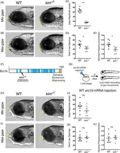
Fig. 1 Erc1b deficiency in kimm533 mutant larvae results in jaw movement defects. A, Minimal, and B, maximal lower jaw displacement from 30 s recordings of jaw movements in 6 dpf WT and kim−/− larvae. Yellow brackets indicate mouth gape. C, Quantification of mean mouth gape magnitude, D, total number of jaw openings, and E, jaw opening frequency (openings per second) in WT (n = 6) and kim−/− (n = 6) larvae. F, Schematic of zebrafish Erc1b protein structure, depicting coiled-coil regions and a C-terminal Rab-binding domain; location of kimm533 mutation indicated. G, Experimental design for WT zebrafish erc1b mRNA injections. H, Minimum, and I, maximum lower jaw displacement of 6 dpf WT erc1b mRNA injected WT and kim−/− larvae. J, Quantification of mean mouth gape magnitude, K, total number of jaw openings, and L, jaw opening frequency in WT (n = 7) and kim−/− (n = 5) larvae injected with WT erc1b mRNA. Symbols indicate individual animals, lines indicate mean with SEM. Mann-Whitney U test (two-tailed) was used for statistical analysis, 95% confidence interval. WT, wild-type. *P < .05, **P < .01