
|
Fig. 2.
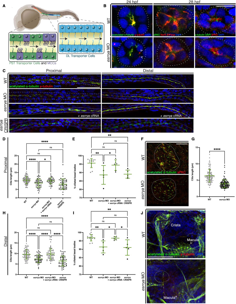
esrrγa is essential for ciliogenesis. (A) Multiciliated cells (MCCs) and monociliated transporter cells in the proximal straight tubule (PST) and distal late (DL). (B,C) IF for indicated markers. Dotted lines indicate the nephron. (D,H) Cilia length. Each dot represents one cilium, and ten cilia were measured per animal. WT n=12, esrrγa MO n=5, esrrγa MO with cRNA n=6, esrrγa crispant n=5. (E,I) Percentage of ciliated basal bodies. Each dot represents one animal. WT n=12, esrrγa MO n=5, esrrγa MO with cRNA n=6, esrrγa crispant n=5. (F) IF for indicated markers in Kupffer's vesicle (KV; outlined with dotted line) at the 10 ss. (G) KV cilia length. Each dot represents a single cilium. WT n=7, esrrγa MO n=7. (J) IF for indicated markers in ear at 4 dpf. Arrowhead denotes macula cilia, dotted line surrounds cristae structures. Data are mean±s.d.. *P<0.05, **P<0.01, ****P<0.0001 (unpaired t-test or one-way ANOVA). ns, not significant. Scale bars: 10 µm (B); 25 µm (F); 50 µm (C,J).