
Image
|
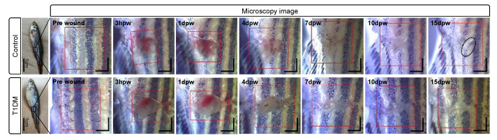
Figure Caption
Figure 3
Bright-field microscopy images of the control (up) and T1DM (down) adult zebrafishes before wounding (pre-wound) and at 3 h, 1 day, 4 days, 7 days, 10 days, and 15 days post-wounding. Red boxes are the scanned areas involving the induced wounds. Scale bars: 1 mm.
Acknowledgments
This image is the copyrighted work of the attributed author or publisher, and
ZFIN has permission only to display this image to its users.
Additional permissions should be obtained from the applicable author or publisher of the image.
Full text @ Bioengineering (Basel)