IMAGE

Figure 3

ID
ZDB-IMAGE-230530-23
Source
Figures for Adrião et al., 2023
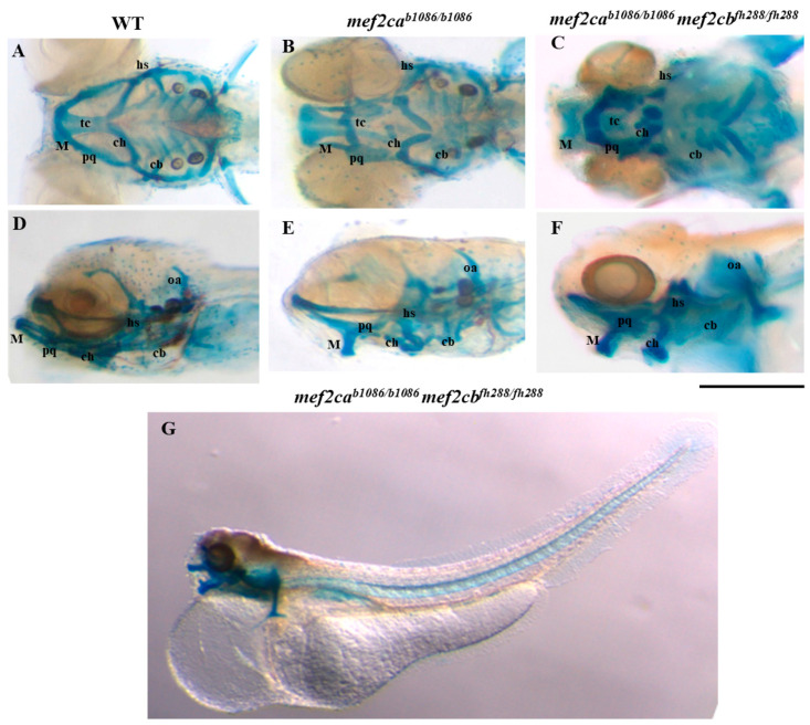
Image
Figure Caption

Figure 3

Craniofacial cartilage phenotypes in WT, mef2ca single-mutant and homozygous double-mutant larvae. The alcian blue staining technique was used to analyse craniofacial cartilaginous phenotype in zebrafish larvae at 5 dpf. (AC) Ventral view of WT, mef2ca single-mutant or homozygous double-mutant larvae. (DF) Lateral view of WT, mef2ca single-mutant or homozygous double-mutant larvae. (G) Lateral view of the whole body of homozygous double-mutant larvae. hs—hyosymplectic cartilage; M—Meckel’s cartilage; ch—ceratohyal; pq—palatoquadrate cartilage; cb—ceratobranchials; oa—occipital arch; tc—trabecula communis. Scale bars: 600 µm.

Acknowledgments
This image is the copyrighted work of the attributed author or publisher, and ZFIN has permission only to display this image to its users. Additional permissions should be obtained from the applicable author or publisher of the image. Full text @ Biomolecules