
|
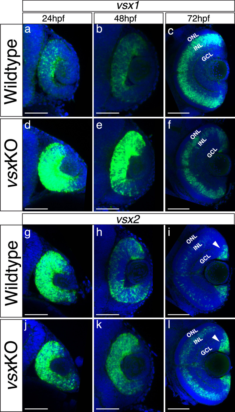
Figure 5—figure supplement 1.
(a-l). Fluorescent in situ hybridization for vsx1 (a-f) and vsx2 (g-l) genes over retina formation in wildtype (a-c, g-i) and vsxKO (d-f) animals (j-l). In wildtype samples, vsx2 is strongly expressed than vsx1 in retinal precursors at earlier developmental stages (a, g) and the opposite effect is observed in INL bipolar cells at later stages (c, i). We found an overexpression of vsx1 at 24hpf (b) and 48hpf (e) and a reduction in the INL expression at 72hpf (c, f) in vsx mutant retinas compared to wildtype. For vsx2, no major differences between wildtype and mutant retinas were detected at 24hpf (g, j), but at 48 and 72hpf we found an increment of vsx2 in the retina and INL, respectively (h, k, i, l). Also, at 72hpf, there is a stronger expression of vsx2 in the ciliary marginal zone (CMZ) compared to WT. hpf: hours post fertilization, ONL: outer nuclear layer, INL: inner nuclear layer, GCL: ganglion cell layer. Scale bar in (a-l): 50 µm.