IMAGE

Figure S1.

ID
ZDB-IMAGE-230523-22
Source
Figures for Cevik et al., 2023
Image
Figure Caption

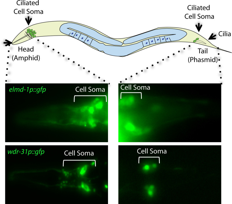
Figure S1. Expression patterns of WDR-31 and ELMD-1.

A total of 60 sensory neurons are distributed in the head (amphid) and tails (phasmid) of C. elegans. The ciliated sensory neurons are displayed in the schematic of C. elegans. The expression of elmd-1promoter::gfp (1,000 kb) and wdr-31promoter::gfp (1,000 kb) were shown in the fluorescence images. The cell of ciliated sensory neurons (cell soma) in the head and tails were displayed in brackets.

Acknowledgments
This image is the copyrighted work of the attributed author or publisher, and ZFIN has permission only to display this image to its users. Additional permissions should be obtained from the applicable author or publisher of the image. Full text @ Life Sci Alliance