
|
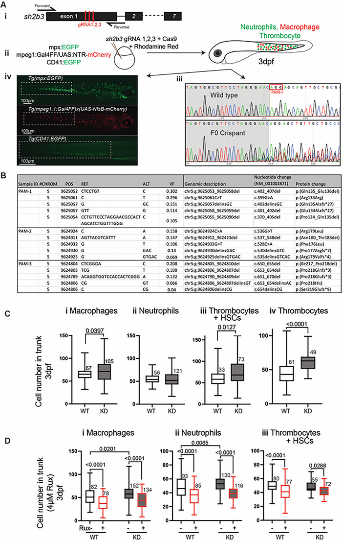
FIGURE 2
Zebrafish sh2b3 loss‐of‐function model. (Ai) CRISPR/Cas9‐mediated mutagenesis to truncate the sh2b3 at exon 1. Red bars indicate the target sites of three designed gRNAs. (Aii) Microinjection of 3x gRNAs into one‐cell stage zebrafish embryo along with Cas9 protein and rhodamine. (Aiii) Sanger sequencing of sh2b3 locus in wild type (top) and F0 crispant (bottom) zebrafish embryos indicating on‐target gene disruption at gRNA‐1 target site (as an example). The red rectangle indicates the PAM sequence for the gRNA‐1. (Aiv) Respectively, EGFP and mCherry expressing neutrophils and macrophages in Tg(mpx:EGFP) and Tg(mpeg1.1:Gal4FF/UAS:NfsB‐mCherry), and EGFP expressing cells (HSCs and thrombocytes) in Tg (CD41:EGFP) 3dpf zebrafish embryos. Dashed white rectangles mark regions in which cells have been quantified. (B) NGS result showing the top 5 most common variants at each gRNA target site. (C) Quantification of macrophages (mpeg1:mCherrypositive), neutrophils (mpx:EGFPpositive), HSCs and thrombocytes (CD41:EGFPpositive) and thrombocytes (mpl:EGFPpositive) in the tail region of 3 dpf sh2b3 knockdown (F0 crispants) compared to wildtype embryos. (D) Quantification of macrophages, neutrophils and