Fig. 3
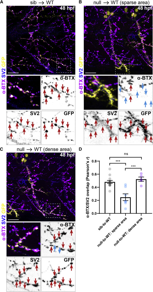
Figure 3. SNRNP70 is cell-autonomously required in motor neurons for neuromuscular assembly (A) A representative image showing transplanted sibling motor neurons derived from a Tg(mnx1:GFP) line innervating a WT host myotome that has been stained with anti-SV2 and α-BTX to mark pre- and post-synaptic sites, respectively. Bottom: inset depicting the apposition (red arrows) of pre- (within the GFP+ motor neurons) and post-synaptic sites within a small region of the myotome. Scale bars, 25 μm. (B and C) Examples of transplanted GFP+ null motor neurons innervating WT host myotomes that have been stained with anti-SV2 and α-BTX. In (B), null GFP+ motor axons terminate in a motor-axon-sparse region of the myotome, whereas in (C), they terminate in a motor-axon-dense region. Bottom: insets depicting the apposition (red arrows) of pre- (within the GFP+ motor neurons) and post-synaptic sites regions of the myotome. Cyan arrows indicate the absence of host AChR clusters adjacent to transplanted GFP+/SV2+ terminals. Scale bars, 25 μm. (D) Quantification of the degree of overlap between SV2 and α-BTX in the three groups of transplanted motor axons. The graph shows mean values ± SEM. ∗∗∗p< 0.001, one-way ANOVA, n = 6 donor/host pairs for sib-to-WT group, n = 3 donor/host pairs for null-to-WT group in three independent experiments. See also Figure S3.
Image
Figure Caption
Acknowledgments
This image is the copyrighted work of the attributed author or publisher, and
ZFIN has permission only to display this image to its users.
Additional permissions should be obtained from the applicable author or publisher of the image.
Full text @ Curr. Biol.

