
|
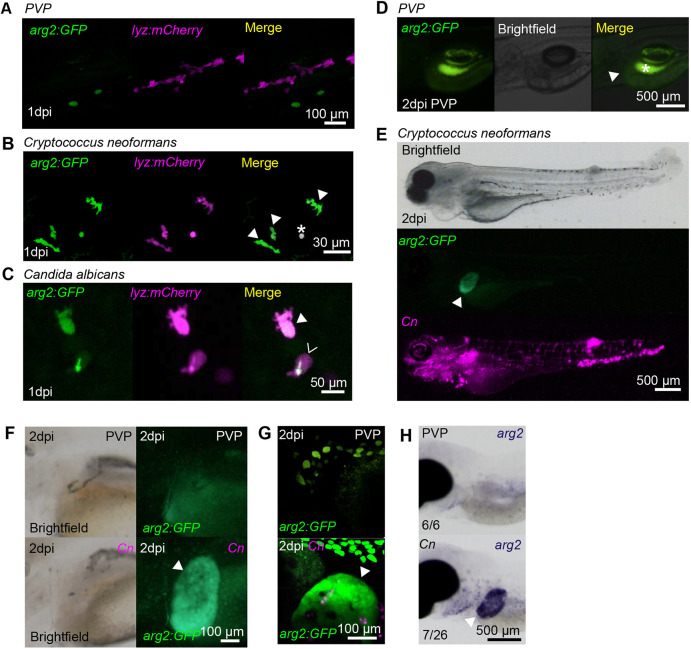
Fig. 4.
Fungal infections upregulate arg2:GFP expression in neutrophils and the liver. (A) Fluorescence confocal micrographs of 2 dpf embryos (arg2:GFP line crossed to the lyz:mCherry line) after PVP mock infection at 1 dpi showing no overlap between GFP and lyz:mCherry expression. (B) Fluorescence confocal micrographs of 1 dpi (2 dpf) embryos (arg2:GFP line crossed to the lyz:mCherry line) after Cryptococcus neoformans infection at 1 dpf showing GFP-positive neutrophils (filled arrowheads). The asterisk indicates a Cryptococcus that has autofluorescence in both channels. (C) Fluorescence confocal micrographs of 1 dpi (2 dpf) embryos (arg2:GFP line crossed to the lyz:mCherry line) after Candida albicans infection at 1 dpf showing a GFP-positive neutrophil (filled arrowhead) and a GFP-negative neutrophil (hollow arrowhead (the green fluorescence signal in this cell is autofluorescence from Candida, which in this instance has survived and formed a hypha). (D) Brightfield and fluorescence micrographs of arg2:GFP larvae at 2 dpi (3 dpf) after PVP injection at 1 dpf. The position of the arg2:GFP-negative liver is shown by the arrowhead. The contrast of the green fluorescence channel has been turned up sufficiently to show gut fluorescence (asterisk), in order to show that the liver is arg2:GFP negative. (E) Brightfield and fluorescence micrographs of 2 dpi (3 dpf) arg2:GFP larvae after Cryptococcus neoformans (Cn) infection at 1 dpf showing arg2:GFP liver-specific expression in an individual with high levels of infection. (F) Brightfield and widefield fluorescence micrographs of 2 dpi (3 dpf) arg2:GFP larvae after PVP mock infection or Cn infection at 1 dpf, showing arg2:GFP liver-specific expression (arrowhead) in the Cn-infected individual. (G) Fluorescence confocal micrographs of 2 dpi (3 dpf) arg2:GFP larvae after PVP mock infection or Cn infection at 1 dpf showing arg2:GFP liver-specific expression (arrowhead) in the Cn-infected individual (arrowhead). (H) Brightfield stereo micrographs of 2 dpi (3 dpf) embryos after PVP or Cn infection at 1 dpf and arg2 whole-mount in situ hybridisation at 3 dpf showing arg2 liver-specific expression (arrowhead) in an infected individual, not present in the PVP-injected larvae. Liver-specific expression of arg2 was observed in n=7/26 Cn-infected larvae performed over three independent experiments.