
|
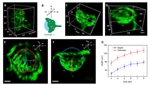
Fig. 7 Development of zebrafish hyaloid vasculature. (A) Typical head vessel structure; red arrow indicates the hyaloid vessel. See Visualization 5. (B) Schematic of the hyaloid basket from ventral-dorsal view. (C–D) Typical hyaloid vessels at 3 dpf and 7 dpf, respectively. See Visualization 6 and Visualization 7. (E) Projection along the lens axis of a typical hyaloid at 5 dpf showing an ellipse shape (red); blue arrows indicate its long axis and short axis, and their mean value is taken as the diameter of the hyaloid basket. (F) Projection along the V-D axial of the hyaloid showing a basket shape. The blue dashed circle indicates the lens, and brown arrows indicate the depth of the basket from the lower edge to the bottom. (G) Diameters (red) and depths (blue) of hyaloid baskets dependent on time show a continuous growth from 3 to 8 dpf. Scale bar: 40 μm for (E) and (F). D: dorsal, V: ventral, N: nasal, T: temporal, La: lens anterior, Lp: lens posterior. Error bars represent the corresponding standard deviation.