
Figure 3
- ID
- ZDB-IMAGE-230430-31
- Genes
- Publication
- Niu et al., 2023 - vwa1 Knockout in Zebrafish Causes Abnormal Craniofacial Chondrogenesis by Regulating FGF Pathway
- All Figures
- Figures for Niu et al., 2023
|
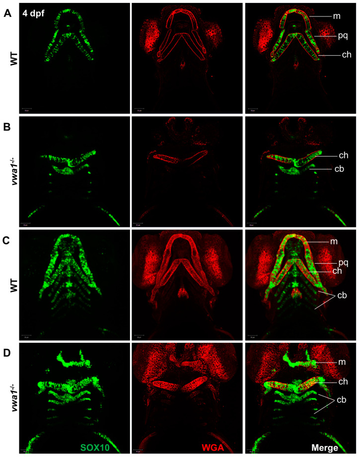
Figure 3
Immunofluorescence assay of WGA and SOX10 reveals a disorganized arrangement of chondrocytes in mutant embryos. (A,B) A WT control embryo at 4 dpf, with single-layer images (A) showing normal craniofacial cartilage morphogenesis and stacked images (B) showing the characteristic “stack of pennies” arrangement of cartilage cells in which the slender cartilage cells assembled with each other to form their own cartilage elements. (C,D) Compared with the WT control group, (C) single-layer and (D) stacked images showed deformities in the overall morphology of the mandibular cartilage of vwa1 mutants, a near absence of Meckel’s cartilage and palatal square cartilage, severely deformed ceratohyal, and smaller size and aspect ratio of many chondrocytes. m, Meckel’s cartilage; pq, palatoquadrate; cb, ceratobranchial; ch, ceratohyal.