Fig. 11
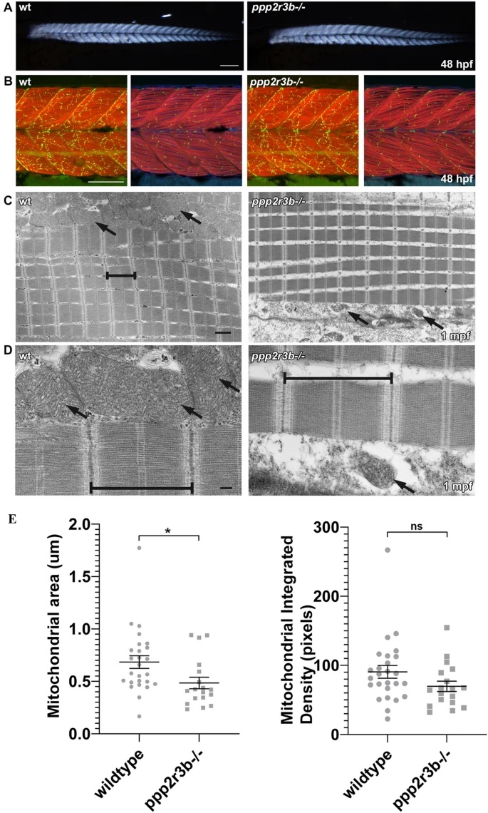
ppp2r3b mutants display abnormal mitochondria formation during adult stages of development, whilst muscle formation remains intact. (A) Skeletal muscle birefringence at 48hpf shows comparable muscle integrity between ppp2r3b mutants and wildtype siblings. Scale bar 200 µm. (B) Muscle fibres and motor neuron synapses appear normal in mutants compared to wildtype siblings at 48hpf, analysed by staining for F-Actin (Red) and Acetylcholine receptors (AChR, green) respectively. Left panels show a z-stack projection of F-Actin with AChR, right panels show a single focal plane of F-Actin/AChR/DAPI. Scale bar 100 µm. (C) Transmission electron micrographs indicate normal sarcomeric assembly (brackets) in juvenile mutants + compared to wildtype controls, however mitochondria (arrows) are noticeably malformed and less abundant. Scale bar 1 µm. (D) High magnification electron micrographs showing detailed images of the sarcomeres (brackets) and mitochondria (arrows) in wildtype compared to mutant adolescent muscle samples. Scale bar 200 nm. (E) Quantification of mitochondrial area adjacent to sarcomeres showed a statistically significant reduction in mutants (t-test) (n = 22 and 17 biological replicates for wild-type and ppp2r3b−/− animals, respectively).

