Fig. 2
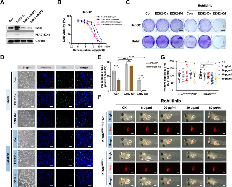
Elevated EZH2 levels lead to antagonism of HCC against FGFR4 inhibitors. A Western blot analysis of EZH2 expression in HepG2 cells transfected with empty vector control (Con), FLAG-EZH2 (EZH2-Ov) and EZH2-siRNAs (EZH2-siRNA1 and 2). B Cell viability of HepG2 cells transfected with empty vector control (Con), FLAG-EZH2 (EZH2-Ov) and EZH2-siRNAs (EZH2-siRNA1 and 2) was evaluated by the CCK-8 following increasing concentrations of Roblitinib treatment for 48 h. Data are presented as mean ± SEM (n = 3). C Crystal violet staining of HepG2 and Huh7 cell lines transfected with empty vector control (Con), FLAG-EZH2 (EZH2-Ov) and EZH2-siRNAs (EZH2-Kd) following Roblitinib treatment for 48 h. Scale bars: 1 cm. D EdU assays of HepG2 cells transfected with empty vector control (Con), FLAG-EZH2 (EZH2-Ov) and EZH2-siRNAs (EZH2-Kd) following Roblitinib treatment for 48 h. Scale bars: 100 μm. E Measurement of the cell numbers in (D). Data are presented as mean ± SEM (n = 3, two-way ANOVA with Sidak’s multiple comparison test, ***p < 0.001, ****p < 0.0001, ns, no significance). F Dose responses of zebrafish KRASG12V+/EZH2+ (top) and KRASG12V+ (bottom) HCC primary tumors treated with Roblitinib for 72 h. Scale bars: 100 μm. G Measurement of the tumor sizes in (F). Data are presented as mean ± SEM (n = 15, two-way ANOVA with Sidak’s multiple comparison test, *p < 0.05, ns, no significance)

